
Single-Nucleotide RNA Pseudouridine (Ψ) & Inosine (I) Modification Seq (BACS-seq)
One chemical conversion is read twice: pseudouridine is written as a protected deletion-type signature and inosine is read as guanosine, so both modifications are mapped and quantified from a single library at single-base resolution.
01What the service reports
Single-Nucleotide RNA Pseudouridine (Ψ) & Inosine (I) Modification Seq (BACS-seq) is an antibody-free, base-resolution method in which a single chemical conversion makes pseudouridine readable as a protected, deletion-type signature and makes inosine readable as guanosine, so both modifications are mapped and quantified together.
Because the two modifications leave different signatures inside the same converted library, one experiment returns paired site maps for pseudouridine and for inosine instead of two separate profiling runs. Each reported site carries a quantified modification ratio rather than an enrichment score.
Arraystar runs BACS-seq as a complete sample-to-data service: RNA sample QC, chemical treatment, library construction, sequencing and bioinformatics analysis. Every call is annotated to the reference transcriptome with genomic coordinates, its host gene and its functional region, and a study can be scoped to mRNA alone or to mRNA together with long non-coding RNA.
02Benefits
- Two modifications from one conversion: pseudouridine and inosine are resolved position by position in the same library rather than in two separately prepared experiments.
- Quantified, not just enriched: conversion-based readouts report a modification ratio at each site, so a call carries a level as well as a position.
- No antibody in the reaction: without immunoprecipitation there is no antibody cross-reactivity to correct for and no pull-down bias to interpret around.
- Every call is annotated: sites are placed on the reference transcriptome with genomic coordinates, host gene and functional region attribution.
- Fitted to downstream questions: site-level output suits work on translation control, transcript stability, coding-region function and gene regulation.
| Service Name | RNA Class | Price |
|---|---|---|
| Single-Nucleotide RNA Pseudouridine (Ψ) & Inosine (I) Modification Seq (BACS-seq) | mRNA | |
| Single-Nucleotide RNA Pseudouridine (Ψ) & Inosine (I) Modification Seq (BACS-seq) | mRNA and lncRNA | |
| Single-Nucleotide RNA Pseudouridine (Ψ) & Inosine (I) Modification Seq (BACS-seq) | rRNA |
03Background and detection principle
In mRNA both pseudouridine and inosine influence how a transcript behaves, so a map is only as useful as its ability to place each modification at a position and give it a level. Enrichment-based profiling reports fragments rather than nucleotides and leaves the two modifications tangled together in the same signal.
BACS-seq separates them chemically. A single conversion step writes pseudouridine as a protected, deletion-type signature while inosine pairs as guanosine during reverse transcription, and unmodified positions read normally. Because both signatures come out of one reaction, the resulting sites are directly comparable instead of being matched across libraries prepared at different times.
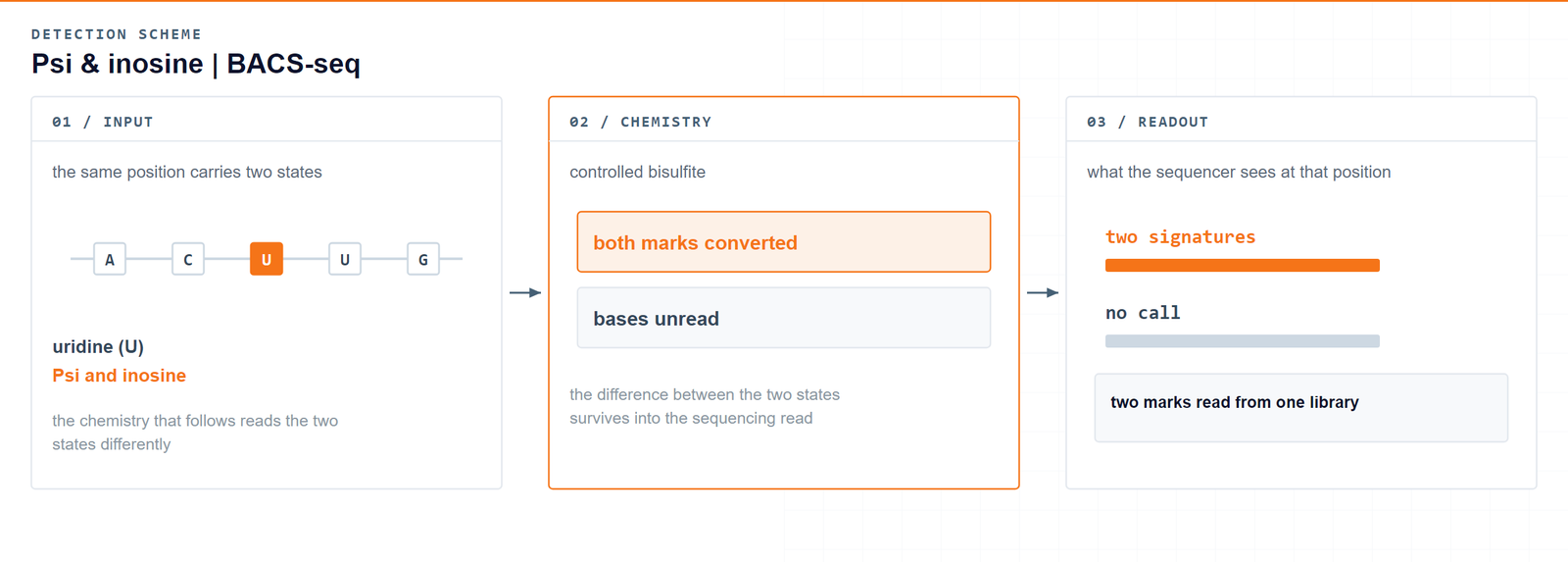
One conversion, two readable signatures: pseudouridine survives as a protected deletion-type signal, inosine reads as guanosine, and unmodified positions read normally.Xu H. et al., Nature Methods 2024 · DOI 10.1038/s41592-024-02439-8
04Workflow
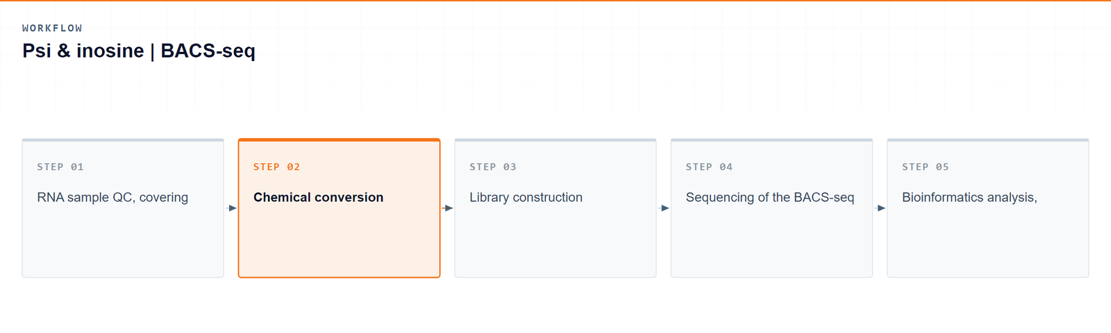
Five steps carry a sample from RNA to single-base pseudouridine and inosine data:
- RNA sample QC, covering integrity and purity before the reaction.
- Chemical conversion of the RNA, which writes the pseudouridine and inosine signatures.
- Library construction from the converted RNA.
- Sequencing of the BACS-seq library.
- Bioinformatics analysis, in which the two signature classes are classified separately and every site is quantified.
05Bioinformatics and deliverables
Reads are aligned and the two signature classes — pseudouridine deletion-type and inosine to guanosine conversion — are called on their own terms, each with a stoichiometry. On top of the site calls the analysis adds motif characterization, transcript-feature distribution, differential modification, functional enrichment and genome-browser visualization.
Deliverables
- Raw pseudouridine and inosine mRNA sequencing data files (FASTQ), formatted for GEO/SRA submission.
- Sample and sequencing QC reports with sorted BAM alignment files and core mapping statistics.
- High-confidence single-base pseudouridine and inosine site tables (XLSX) listing coordinates, host genes and stoichiometry.
- Single-base pseudouridine and inosine transcript-feature distribution plots and consensus motif logos (PDF/PNG).
- Single-base differential pseudouridine and inosine tables (XLSX) with fold change and significance.
- GO enrichment reports for pseudouridine- and inosine-modified genes (HTML/PDF/PNG).
- Genome-browser signal tracks (bigWig/bedGraph) together with a structured project report.
06Research applications
- Transcriptome-wide maps: pseudouridine and inosine sites are resolved across protein-coding transcripts and, where the study is scoped that way, across lncRNAs as well.
- Translation and transcript stability: site-level modification data can be set against how efficiently an mRNA is translated and how long it persists.
- Coding-region and UTR function: sites are localized within CDS, UTR and splice-adjacent regions, so positional effects can be examined rather than assumed.
- Disease-oriented work: differentially modified mRNA sites can be carried forward as candidate biomarkers.
- Cross-modification comparison: because both signatures come from the same conversion, the two site maps can be compared directly within one dataset.
07Sample requirements
Storage. Cells and tissues: preserve in TRIzol or an RNA stabilisation solution, snap freeze in liquid nitrogen and store at −80 °C. RNA: resuspend in ethanol or RNase-free ultrapure water, store at −80 °C and avoid repeated freeze–thaw cycles.
Shipping. Place the sample in a 1.5 mL RNase-free microcentrifuge tube, seal it with parafilm or a cap lock, and ship on dry ice with adequate insulation.
| Sample type | Notes |
|---|---|
| Whole blood | EDTA tubes only; heparin is not compatible with the downstream procedure. |
| Cultured cells | Cell pellets are preferred. |
| Tissue | Fresh or frozen specimens; avoid necrotic material. |
| Total RNA | OD 260/280 ≥ 1.8 and RIN ≥ 7, with no visible degradation. Submit at least 10 µg total RNA. |
08FAQ
What comes back per modification from a BACS-seq run?
A table of single-base sites for pseudouridine and a table of single-base sites for inosine, each with genomic coordinates, the host gene, the functional region and a quantified modification ratio. Motif, distribution, differential and enrichment analyses are built on top of those tables.
Are both modifications really called from one library?
Yes. One conversion step writes the pseudouridine signature and the inosine signature into the same converted RNA, and the two classes are then classified separately during analysis. Nothing has to be split into two experiments for the two modifications to be read out.
How is this different from an antibody-based enrichment assay?
Antibody-based profiling enriches fragments, which caps resolution at the fragment and folds an antibody’s binding preference into the signal. BACS-seq converts the RNA chemically, so calls rest on conversion signatures rather than on capture, and each site arrives with a stoichiometry.
Which transcript scopes can I order?
Two: modification profiling on mRNA, or on mRNA together with long non-coding RNA. Both transcript types are analyzed within a single experiment, so a study that spans coding and non-coding transcripts does not have to be run as two projects.
How are the reported sites annotated?
Each site is placed on the reference transcriptome with its genomic coordinates, its host gene and its functional region attribution, so a call can be interpreted in the context of the transcript it belongs to.
09References
- Xu H, Kong L, Cheng J, et al. Absolute quantitative and base-resolution sequencing reveals comprehensive landscape of pseudouridine across the human transcriptome. Nature Methods 2024;21(11):2024–2033. DOI: 10.1038/s41592-024-02439-8