
Single-Nucleotide RNA m1A Modification Seq (m1A-Quant-seq)
An engineered reverse transcriptase copies straight through m1A and leaves a characteristic mismatch behind it, so the mutation rate at a position reports modification stoichiometry instead of ending the read.
01What the service reports
Single-Nucleotide RNA m1A Modification Seq (m1A-Quant-seq) reports where m1A occurs in a transcript and what fraction of the RNA is modified at each of those positions, using an engineered reverse transcriptase that reads through the modified base and records it as a reproducible mismatch.
The enzyme does not stall when it meets m1A. It continues through the site and introduces a characteristic mutation signature, mostly a T in the sequencing read, at exactly that position. Counting how often the signature appears turns the mismatch rate into a modification percentage, so position and level are obtained from the same read pile.
Arraystar offers the service end to end: RNA QC, library construction, sequencing, bioinformatics and annotation, and a structured project report. Each run also carries a demethylated background-control aliquot, which is what allows the signature to be read as m1A rather than as general misincorporation.
02Benefits
- Near single-base precision: individual m1A positions are resolved, unlike the peak-level resolution of antibody enrichment.
- Absolute stoichiometry: the share of m1A at each site is calculated from the ratio of mutation signatures.
- Read-through design: the engineered reverse transcriptase is not stopped by m1A, so reads are not lost to premature termination.
- Workable with limited material: the chemistry is designed for studies where only small amounts of RNA are available, such as rare clinical specimens.
- No antibody bias: without immunoprecipitation there is no non-specific binding, no antibody cross-reactivity and batch-to-batch antibody variation are not sources of variation in this assay.
| Service Name | RNA Class | Price |
|---|---|---|
| Single-Nucleotide RNA m1A Modification Seq (m1A-Quant-seq) | mRNA | |
| Single-Nucleotide RNA m1A Modification Seq (m1A-Quant-seq) | mRNA and lncRNA | |
| Single-Nucleotide RNA m1A Modification Seq (m1A-Quant-seq) | rRNA |
03Background and detection principle
Classic antibody-based m1A mapping, such as m1A-MeRIP-seq, reports peaks rather than positions and suffers from severe reverse-transcription arrest at the modified base. Modified fractions therefore cannot be quantified directly, which leaves a blind spot in m1A research.
m1A-Quant-seq addresses both problems at once. An engineered reverse transcriptase is combined with optimized reaction conditions so that the enzyme copies through m1A and converts the site into a precise, reproducible mismatch signature instead of a truncation. The signature is then quantified against a demethylated background control, producing absolute stoichiometry at single-nucleotide resolution from low-input RNA.
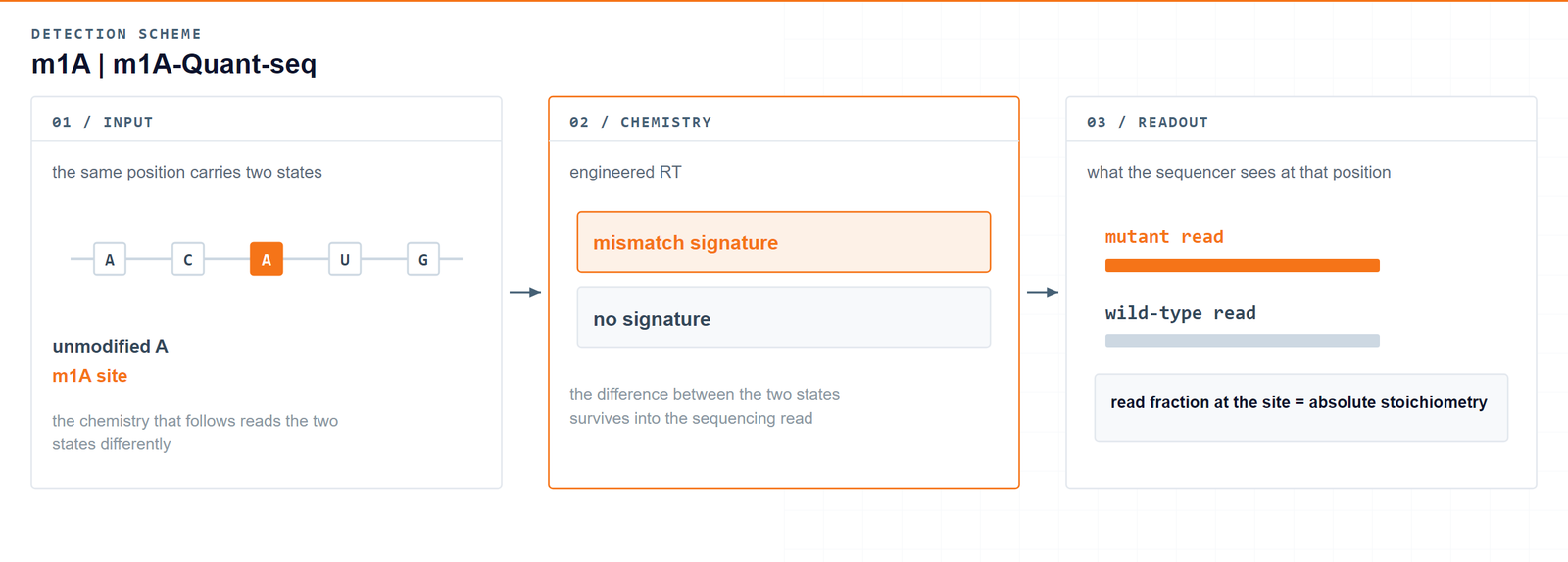
Reading through the modification rather than stopping at it: the mismatch left behind at a base is what turns m1A into a number.Li X. et al., Molecular Cell 2017 · DOI 10.1016/j.molcel.2017.10.019
04Workflow
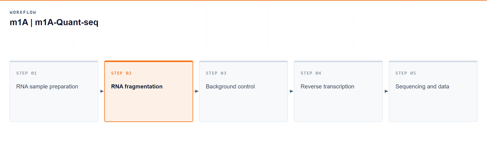
Five stages take a sample from total RNA to quantified m1A sites:
- RNA sample preparation: purified total RNA is selected for mRNA by poly(A) selection.
- RNA fragmentation to the fragment size the library needs.
- Background control preparation: one aliquot is demethylated with AlkB, while a second aliquot is left untreated.
- Reverse transcription: both aliquots are converted into sequencing libraries with the engineered reverse transcriptase.
- Sequencing and data analysis: reads are mapped, and m1A is called from the mutation signature with background correction applied.
05Bioinformatics and deliverables
The pipeline maps m1A at single-nucleotide resolution, quantifies the m1A methylome and compares modification between conditions, which is what makes downstream biological and biomarker questions addressable. Most tables and figures open in standard office software, and the raw datasets are provided for further computational work or for deposition.
Deliverables
- Raw m1A mRNA sequencing data files (FASTQ).
- Sample and sequencing QC reports, together with sorted BAM alignments and core mapping statistics.
- High-confidence single-base m1A site tables (XLSX) with genomic coordinates, host genes and absolute modification stoichiometry (m1A%), plus codon position, the encoded amino acid and an evolutionary conservation score for each site.
- Transcript feature distribution figures and motif logos (PDF/PNG).
- Differential methylation tables (XLSX).
- GO enrichment reports (HTML/PDF/PNG).
- Genome-browser-compatible tracks (bigWig/bedGraph) and a structured project report.
Optional advanced analysis
- Transcript expression correlation, relating m1A levels to the expression of the modified transcripts.
- Translation efficiency inference, exploring how m1A levels relate to how efficiently target mRNAs are translated.
- RNA stability inference, evaluating how m1A levels relate to the half-life of target mRNAs.
- Conserved m1A sites across species, flagging positions that may carry conserved functions.
- Global distribution heatmaps and scatterplots linking transcript abundance or half-life to modification level.
06Research applications
m1A is a dynamic post-transcriptional modification that matters for RNA stability, structural folding and translational control. Carrying a positive charge under physiological conditions, it blocks standard Watson-Crick pairing and forces structural changes in mRNA, tRNA and rRNA; dysregulation of m1A pathways has been implicated in cancers, metabolic syndromes and neurological disorders.
- Oncogenesis — biomarker discovery: tumour biopsies profiled against healthy margins yield absolute stoichiometry, so high-percentage m1A sites can be used as candidates for biomarker exploration in disease staging. [1, 2, 3]
- Oncogenesis — metabolic rewiring: work in hepatocellular carcinoma and glioblastoma links altered m1A levels on metabolic transcripts to the translation of glycolytic enzymes, which supports tumour adaptation to hypoxia. [1, 2]
- Oncogenesis — splicing variation: single-nucleotide resolution places sites directly at intron-exon junctions, which allows aberrant m1A masking and the resulting tumour-specific neoantigens to be examined. [1, 2]
- Neurological disorders — neural stem cell differentiation: m1A fluctuations can be followed through early embryonic neurogenesis, where shifts in stoichiometry on neurodevelopmental transcripts accompany the choice between self-renewal and differentiation.
- Neurological disorders — neurodegenerative mechanisms: in Alzheimer's and Parkinson's disease models, sites can be mapped on transcripts governing mitochondrial function and proteasome clearance, connecting loss of m1A homeostasis to neurotoxicity and synaptic loss. [1, 2, 3]
- Immunological and viral epitranscriptomics — viral lifecycle mapping: applied to viral genomes such as HIV-1, Zika or influenza, the method identifies structural modifications that help viral RNA escape host pattern-recognition receptors. [1]
- Immunological and viral epitranscriptomics — immune evasion: quantitative mapping shows how low-level m1A changes on host interferon-stimulated genes modulate the kinetics of the antiviral response. [1]
07Sample requirements
Purified total RNA is the preferred input. RNA extraction from cells, tissue or blood can also be arranged as part of the project; if the amount of material you can obtain is limited, tell us what is realistically available so the options can be discussed.
Storage. Cells and tissues: preserve in TRIzol (cells or tissues) or RNAlater (tissue) and store at −80 °C. RNA: resuspend in ethanol or RNase-free ultrapure water, store at −80 °C and avoid repeated freeze–thaw cycles.
Shipping. Place the sample in a 1.5 mL RNase-free microcentrifuge tube, seal it with parafilm or a cap lock, and ship on dry ice with adequate insulation.
| Sample type | Notes |
|---|---|
| Whole blood | EDTA tubes only; heparin is not compatible with the downstream procedure. |
| Cultured cells | Cell pellets are preferred. |
| Tissue | Fresh or frozen specimens; avoid necrotic material. |
| Total RNA | OD 260/280 ≥ 1.8 and RIN ≥ 7, with no visible degradation. Submit at least 10 µg total RNA. |
08FAQ
Why measure m1A stoichiometry instead of only presence?
m1A levels move with condition and with the enzyme that writes or removes the mark. A position that changes in occupancy, not merely in detectability, is the observation that connects a modification call to a regulatory question on mRNA or long non-coding RNA.
Can m1A-Quant-seq call m1A at single-base resolution?
Yes. The modification is detected through the signature it induces at that base during reverse transcription, so sites are localised and quantified at individual nucleotide positions. A demethylated aliquot is processed alongside the sample as a background control.
How does this service compare with m1A-MeRIP-seq?
Antibody-based m1A mapping reports peaks of roughly 100–200 nt and suffers from reverse-transcription arrest at the modified base, which rules out absolute quantification. m1A-Quant-seq reads through the site and quantifies the signature instead, so sites are resolved individually and modification levels are measured rather than inferred.
Which research applications fit this readout?
Site-level m1A maps support work on transcript fate, translation control and disease-associated remodelling of the methylome. Because each call carries a modification ratio, candidates can be ranked for follow-up rather than treated as a binary list.
What does a site list make possible in a study?
Annotated positions on mRNA and long non-coding RNA, with modification ratios and region labels, are the starting set for condition contrasts, motif checks and focused validation of the sites that move. Differential tables and browser tracks are part of the same package.
09References
- Li X, Xiong X, Zhang M, et al. Base-resolution mapping reveals distinct m1A methylome in nuclear- and mitochondrial-encoded transcripts. Molecular Cell 2017;68(5):993–1005.e9. DOI: 10.1016/j.molcel.2017.10.019
- Kvolik Pavić A, Čonkaš J, Mumlek I, et al. Clinician's guide to epitranscriptomics: an example of N1-methyladenosine (m1A) RNA modification and cancer. Life 2024;14(10):1230. DOI: 10.3390/life14101230
- Pajdzik K, Lyu R, Dou X, et al. Chemical manipulation of m1A mediates its detection in human tRNA. RNA 2024;30(5):548–559. DOI: 10.1261/rna.079966.124