
Single-Nucleotide RNA m6A Modification Seq (CAM-seq)
A chemical, antibody-free route to absolute m6A quantification: cooperative catalysis deaminates unmodified adenosine to inosine while m6A resists, so every modified site is read out from the A-to-G signal at single-base resolution.
01What the service reports
Single-Nucleotide RNA m6A Modification Seq (CAM-seq) is a chemical m6A sequencing method in which cooperative catalysis selectively deaminates unmodified adenosine to inosine while m6A resists, so each modified site is called from an A-to-G signature at single-base resolution.
A carbonyl organocatalyst and a Lewis acid (boric acid) act together under mild, near-neutral conditions. Unmodified adenosine is converted to inosine and is read as guanine during reverse transcription; m6A stays intact and is read as adenine. Because the chemistry distinguishes modified from unmodified bases directly, the readout carries both the position of a site and its modification ratio.
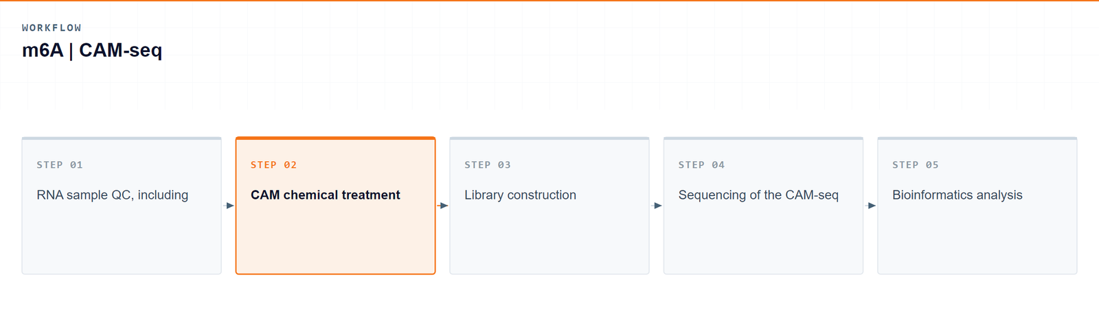
Arraystar runs CAM-seq as a complete laboratory workflow: RNA QC, the CAM chemical treatment, library construction, sequencing and bioinformatics analysis. Two transcript scopes are available, so a study can profile mRNA alone or mRNA together with long non-coding RNA in the same experiment.
02Benefits
- Single-base resolution: each m6A site is pinpointed by its own A-to-G signature rather than inferred from a peak region.
- Absolute stoichiometry: the A/G ratio at a site reports the modification ratio, not only an enrichment score.
- Mild near-neutral chemistry: a reversible kethoxal guanine-protecting group helps keep the RNA intact through the reaction.
- Antibody-free: there is no antibody step, so the profile is not shaped by antibody binding preference; motif calling is not restricted to a canonical motif, so sites outside the standard motif are not systematically missed.
- Low background: optimized reverse-transcription conditions keep background signal low so that calls remain reliable.
| Service Name | RNA Class | Price |
|---|---|---|
| Single-Nucleotide RNA m6A Modification Seq (CAM-seq) | mRNA | |
| Single-Nucleotide RNA m6A Modification Seq (CAM-seq) | mRNA and lncRNA | |
| Single-Nucleotide RNA m6A Modification Seq (CAM-seq) | rRNA |
03Background and detection principle
Antibody-based m6A profiling, such as MeRIP-seq, enriches fragments rather than resolving positions, so its resolution is limited and its signal carries the motif preference of the antibody. Earlier chemical deamination approaches reach base resolution but rely on acidic conditions that damage RNA and push input requirements up.
CAM-seq works around both limits with cooperative catalysis. A carbonyl organocatalyst and boric acid selectively deaminate unmodified adenosine to inosine at mild, near-neutral pH, while m6A resists the reaction. A reversible kethoxal group protects guanine during the treatment, and the optimized reaction and reverse-transcription conditions keep background signal low. The result is a quantitative readout of m6A at true single-base resolution.
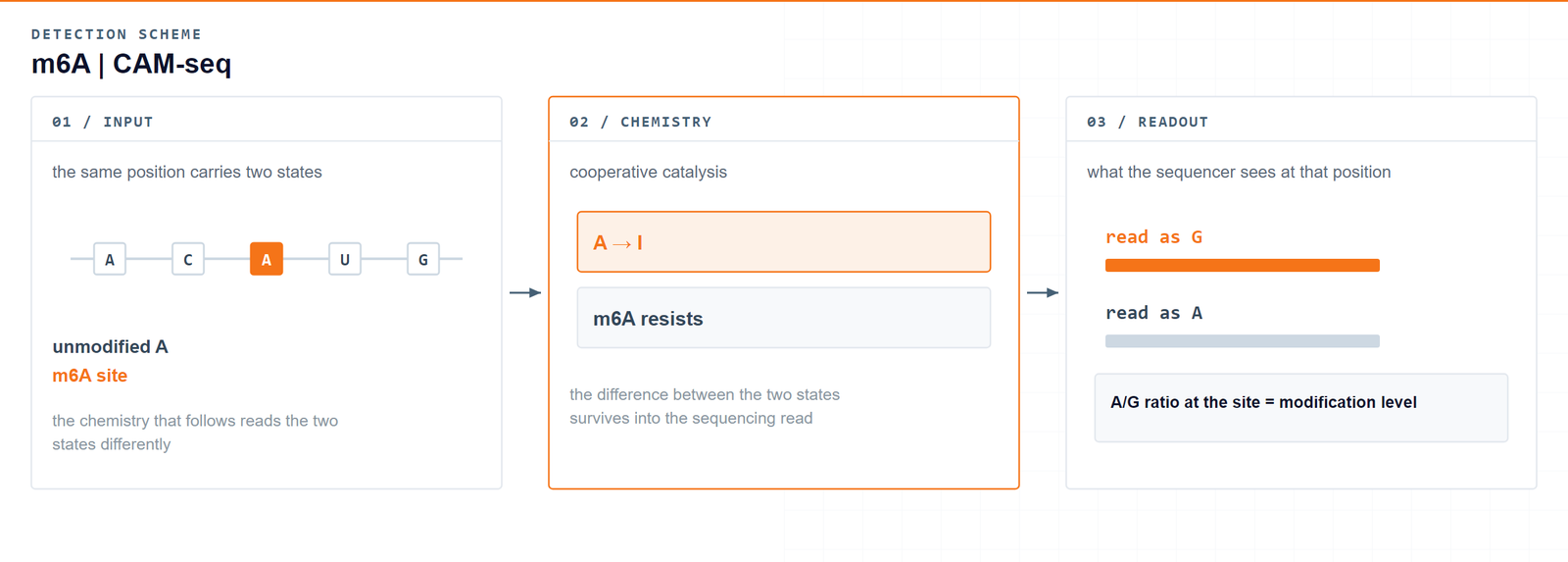
Cooperative catalysis deaminates unmodified adenosine while m6A resists — so the position and the level of a modification come out of the same signal.Wang P. et al., Nature Chemistry 2025 · DOI 10.1038/s41557-025-01801-3
04Workflow
Five steps take a sample from RNA to single-base m6A data:
- RNA sample QC, including integrity and purity checks before the reaction.
- CAM chemical treatment under mild, near-neutral conditions with the kethoxal protecting group.
- Library construction from the treated RNA.
- Sequencing of the CAM-seq library.
- Bioinformatics analysis to call m6A sites and quantify their stoichiometry.
05Bioinformatics and deliverables
Reads are mapped to the reference genome, A-to-G conversion signatures are detected, and m6A stoichiometry is quantified at single-nucleotide resolution. The analysis set covers motif characterization (including GAC, AAC and non-canonical motifs), metagene distribution, differential methylation, functional enrichment and genome-browser visualization.
Deliverables
- Raw m6A mRNA sequencing data files (FASTQ), formatted for GEO/SRA submission.
- QC reports and sorted, indexed BAM alignment files with core mapping statistics.
- High-confidence single-base m6A site tables (XLSX) with genomic coordinates, host genes and modification stoichiometry.
- Single-base m6A transcript feature distribution plots and consensus motif logos (PDF/PNG).
- Single-base differential m6A methylation tables (XLSX) with fold change and significance.
- GO enrichment reports for m6A-modified genes (HTML/PDF/PNG).
- Genome-browser signal tracks (bigWig/bedGraph) and a structured project report.
06Research applications
- Plant and crop research: cross-species single-base m6A maps, including published sets of 111,121 Arabidopsis and 141,990 maize sites, extend epitranscriptomic work from model plants into crops.
- Neuroscience: single-base m6A landscapes of brain tissue and neuronal transcripts support studies of m6A in neural function and disease.
- Cancer research: site-level m6A stoichiometry compared between tumour and normal samples helps identify dysregulated methylation and candidate biomarkers.
- Stem cell and developmental biology: resolves m6A dynamics at individual sites during cell fate commitment and tissue development.
- Gene regulation and systems biology: saturated m6A maps with quantified stoichiometry allow gene-level comparisons across genes, pathways and conditions.
07Sample requirements
Storage. Cells and tissues: preserve in TRIzol or an RNA stabilisation solution, snap freeze in liquid nitrogen and store at −80 °C. RNA: resuspend in ethanol or RNase-free ultrapure water, store at −80 °C and avoid repeated freeze–thaw cycles.
Shipping. Place the sample in a 1.5 mL RNase-free microcentrifuge tube, seal it with parafilm or a cap lock, and ship on dry ice with adequate insulation.
| Sample type | Notes |
|---|---|
| Whole blood | EDTA tubes only; heparin is not compatible with the downstream procedure. |
| Cultured cells | Cell pellets are preferred. |
| Tissue | Fresh or frozen specimens; avoid necrotic material. |
| Total RNA | OD 260/280 ≥ 1.8 and RIN ≥ 7, with no visible degradation. Submit at least 3 µg total RNA. |
08FAQ
Which sample types are compatible with CAM-seq?
Total RNA extracted from cells, tissues or blood, including cultured cell pellets and frozen tissue specimens. If you are unsure whether a particular sample type or preparation is suitable, contact technical support before submission.
Does CAM-seq resolve m6A at single-base resolution?
Yes. Unmodified adenosine is deaminated to inosine and read as guanine, while m6A is read as adenine. That A-to-G signature locates each site at single-base resolution and gives its modification ratio at the same time.
How does CAM-seq differ from antibody-based m6A methods?
Antibody-based methods such as MeRIP-seq work by fragment enrichment, which limits resolution and carries the motif preference of the antibody. CAM-seq is antibody-free and converts unmodified bases chemically, so detection does not depend on an antibody’s binding preference.
How does CAM-seq compare with other chemical deamination methods?
CAM-seq uses cooperative catalysis — a carbonyl organocatalyst paired with boric acid — under mild, near-neutral conditions with a reversible kethoxal protecting group, and it works from total RNA in a complete sample-to-data workflow.
Can the same experiment cover lncRNA as well?
Yes. Two transcript scopes are offered: modification profiling on mRNA, or on mRNA together with long non-coding RNA. Both transcript types are analyzed in one experiment rather than split into two projects.
09References
- Wang P, Ye C, Zhao M, Jiang B, He C. Small-molecule-catalysed deamination enables transcriptome-wide profiling of N6-methyladenosine in RNA. Nature Chemistry 2025. DOI: 10.1038/s41557-025-01801-3
- Garcia-Campos MA, et al. Deciphering the “m6A code” via antibody-independent quantitative profiling. Cell 2019;178:731–747. DOI: 10.1016/j.cell.2019.06.013