
Single-Nucleotide RNA m7G & m3C Modification Seq (AlkAniline-Seq)
A modified base is a weak point in the chain. Heat and alkali take the base off and leave an abasic site; aniline cuts there and leaves a 5′-phosphate behind, and that phosphate is the only entry point an adapter can use. The cut becomes the coordinate, and one library carries m7G and m3C at the same time. The maps on tRNA and rRNA are the reproducible core; positions on mRNA and lncRNA are exploratory.
01What the service reports
Single-Nucleotide RNA m7G Modification Seq (AlkAniline-Seq) is a chemistry-based sequencing method that reports m7G and m3C at single-nucleotide resolution: heat under alkaline conditions removes the modified base and leaves an abasic site, aniline cuts the chain there, and the 5′-phosphate created at the next residue directs adapter ligation.
Each called position carries a site level, so a modification present on only part of the RNA pool keeps an intermediate value instead of being rounded to a yes or no call. Sites are annotated with the RNA they were read on, the transcript they belong to and the structural region they sit in, and m7G and m3C are reported as two separate lists.
Arraystar runs the route end to end, from RNA QC and the cleavage chemistry through library construction and sequencing to bioinformatics, and returns the site tables for the sample set that was submitted.
02Benefits
- Two marks in the same assay: m7G and m3C are read together, so the two site lists can be compared directly.
- Single-base resolution: a site is the nucleotide where the chemistry stopped, not the center of an enriched interval.
- Positive selection, low background: only ends that carry the new 5′-phosphate enter the library, which is what keeps the reaction background low.
- Chemistry rather than affinity: no antibody takes part, so a binding preference does not shape the site list.
- No RNA-class fractionation: total RNA enters the reaction as it is, so no size or tRNA fractionation is done before the chemistry.
- Annotated output: every site is placed on its host transcript with its RNA class and structural region.
| Service Name | Price |
|---|---|
| Single-Nucleotide RNA m7G Modification Seq (AlkAniline-Seq) — mRNA | |
| Single-Nucleotide RNA m7G Modification Seq (AlkAniline-Seq) — mRNA and lncRNA |
03Background and detection principle
Two modified pyrimidine and purine bases behave in a way that unmodified residues do not: the bond between the base and the ribose is labile. m7G carries a positive charge on the ring nitrogen, and m3C breaks down through the same route, so a combination of heat and alkaline conditions is enough to detach the base and leave an abasic site in the chain. The same chemistry reaches dihydrouridine and 5-hydroxycytidine, which is why the method is described as reading a family of labile-bond marks rather than a single modification.
AlkAniline-Seq builds the library outwards from that abasic site. Aniline drives β-elimination, the backbone is cut, and a 5′-phosphate remains on the residue next to the modified base. Ligation then accepts only those ends, so the reaction positively selects the fragments that carry a mark instead of competing with the unmodified majority of the sample. The marks are separated after alignment: a cut read on a guanosine reports m7G, a cut read on a cytidine reports m3C.
The same principle explains where the maps are strongest. The method was validated on bacterial, yeast and human cytoplasmic and mitochondrial tRNAs and rRNAs from total RNA, and those are the RNA classes in which the site catalog is most complete. Transcript-level positions are reported for the molecules that reach sufficient coverage in the sample, which is a question of depth and of how much of the mark that RNA class actually carries.
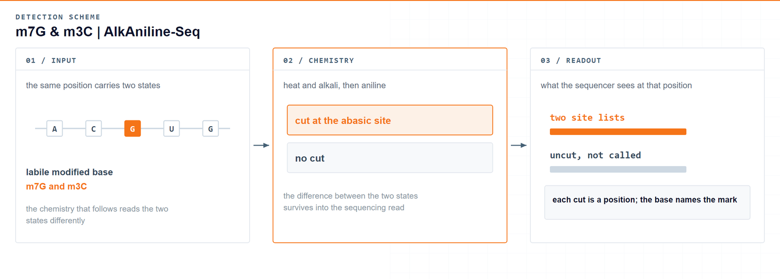
The proposed approach eschews conventional RNA sequencing chemistry and rather exploits the generation of abasic sites and subsequent aniline cleavage. The newly generated 5′-phosphates are used as unique entry for ligation of an adapter in library preparation.Marchand V. et al., Angewandte Chemie International Edition 2018 · DOI 10.1002/anie.201810946
04Workflow
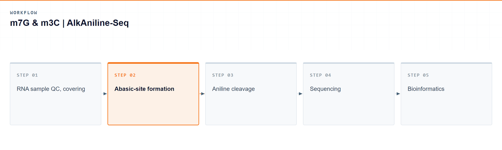
Five steps take a sample from RNA to a single-base m7G and m3C table:
- RNA sample QC, covering integrity and purity.
- Abasic-site formation under heat and alkaline conditions, where the labile base leaves its ribose, followed by dephosphorylation of pre-existing 5′-phosphates.
- Aniline cleavage of the chain, followed by adapter ligation at the new 5′-phosphate ends.
- Sequencing of the AlkAniline-Seq library on an Illumina platform.
- Bioinformatics: alignment, cut-position scoring, and annotation and quantification of the two site lists.
05Bioinformatics and deliverables
Reads are aligned and cut positions are scored nucleotide by nucleotide, with guanosine positions reported as m7G and cytidine positions annotated as m3C at known m3C sites (ho5C is not assigned from the parental base alone). The analysis adds the host transcript and structural region of each site, site-level comparison between sample groups, and genome-browser visualization of the cut profile.
Deliverables
- FASTQ data for every library, formatted ready for GEO/SRA submission.
- A QC report together with sorted BAM alignments and core mapping statistics.
- Single-base m7G site tables (XLSX) with transcript coordinates, host gene and site level.
- Single-base m3C site tables (XLSX) with transcript coordinates, host gene and site level.
- Site distribution figures by RNA class and by structural region (PDF/PNG).
- Differential modification tables (XLSX) with fold change and significance.
- Genome-browser track files (bigWig/bedGraph) and a structured project report.
06Research applications
- Reading two marks together: ask whether an m7G change in a sample is accompanied by an m3C change, from the same preparation and the same run.
- Writer-enzyme studies: follow how METTL1/WDR4 loss or gain shifts m7G positions, and how METTL6 and METTL2A/2B shape the m3C set.
- Translation and codon use: relate a site-level change on a tRNA family to ribosome occupancy and to the translation of the codons that family decodes.
- Cancer and disease models: compare site profiles between tumour and normal material or across disease models in which these writers are dysregulated.
- Cross-method work: set the chemical map beside antibody-based enrichment or beside another base-resolution method run on the same samples.
07Sample requirements
Storage. Cells and tissues: preserve in TRIzol or an RNA stabilisation solution, snap freeze in liquid nitrogen and store at −80 °C. RNA: resuspend in ethanol or RNase-free ultrapure water, store at −80 °C and avoid repeated freeze–thaw cycles.
Shipping. Place the sample in a 1.5 mL RNase-free microcentrifuge tube, seal it with parafilm or a cap lock, and ship on dry ice with adequate insulation.
| Sample type | Notes |
|---|---|
| Whole blood | EDTA tubes only; heparin is not compatible with the downstream procedure. |
| Cultured cells | Cell pellets are preferred. |
| Tissue | Fresh or frozen specimens; avoid necrotic material. |
| Total RNA | OD 260/280 ≥ 1.8 and RIN ≥ 7, with no visible degradation. Submit at least 10 µg total RNA. |
08FAQ
Which modified nucleotides does the chemistry read?
The reaction targets the labile bond between base and ribose. Its two main outputs are m7G and m3C, and the same route reaches dihydrouridine and 5-hydroxycytidine, which are reported when they are present in the sample rather than as a separate service.
Why does this readout not need an antibody?
Because the library is built from the cut. Only ends that carry the newly created 5′-phosphate are ligated to adapters, so the reaction itself selects the marked fragments. That positive selection is what gives the method its low background, and it means no binding preference is imposed on the site list.
How far can the m7G map be extended beyond tRNA and rRNA?
Reports differ, and the difference is worth knowing. Methods that reduce the base chemically before sequencing have mapped m7G on tRNA and rRNA but found no internal m7G in yeast or E. coli mRNA, while other studies report internal m7G in mammalian mRNA and in miRNAs. The maps on tRNA and rRNA are the reproducible core; transcript-level positions depend on the sample, the depth and the method.
What is the evidence for m3C outside tRNA?
m3C in tRNA is long established, and a cleavage-based study of human cells found tRNAs to be the predominant m3C species, with no comparable signal in mRNA or other non-coding RNA. An antibody-based study later reported m3C in mRNA and lncRNA, enriched in 3′ UTRs. A project that cares about this question should size the depth for it.
How is a site quantified?
At every position the pipeline relates the reads starting immediately 3′ of the cut to the coverage at that position and reports the result as a site level. A position modified on part of the RNA pool therefore returns an intermediate value, which is what makes comparisons between groups meaningful.
09References
- Marchand V, Ayadi L, Ernst FGM, et al. AlkAniline-Seq: profiling of m7G and m3C RNA modifications at single nucleotide resolution. Angewandte Chemie International Edition 2018;57(51):16785-16790. DOI: 10.1002/anie.201810946
- Marchand V, Bourguignon-Igel V, Helm M, Motorin Y. Mapping of 7-methylguanosine (m7G), 3-methylcytidine (m3C), dihydrouridine (D) and 5-hydroxycytidine (ho5C) RNA modifications by AlkAniline-Seq. Methods in Enzymology 2021;658:25-47. DOI: 10.1016/bs.mie.2021.06.001
- D'Ambrosi S, Garcia-Vilchez R, Kedra D, et al. Global and single-nucleotide resolution detection of 7-methylguanosine in RNA. RNA Biology 2024;21(1). DOI: 10.1080/15476286.2024.2337493
- Zhang LS, Liu C, Ma H, et al. Transcriptome-wide mapping of internal N7-methylguanosine methylome in mammalian mRNA. Molecular Cell 2019;74(6):1304-1316. DOI: 10.1016/j.molcel.2019.03.036
- Pandolfini L, Barbieri I, Bannister AJ, et al. METTL1 promotes let-7 microRNA processing via m7G methylation. Molecular Cell 2019;74(6):1278-1290. DOI: 10.1016/j.molcel.2019.03.040
- Enroth C, Poulsen LD, Iversen S, Kirpekar F, Albrechtsen A, Vinther J. Detection of internal N7-methylguanosine (m7G) RNA modifications by mutational profiling sequencing. Nucleic Acids Research 2019;47(20):e126. DOI: 10.1093/nar/gkz736
- Cui J, Liu Q, Sendinc E, Shi Y, Gregory RI. Nucleotide resolution profiling of m3C RNA modification by HAC-seq. Nucleic Acids Research 2021;49(5):e27. DOI: 10.1093/nar/gkaa1186