
Single-Nucleotide RNA m7G Modification Seq (m7G-Quant-seq)
Internal m7G is hard to separate from the cap, and that is what this method solves: a targeted chemical conversion treats the two differently, so each internal site is recorded as its own reverse-transcription signature and read at base resolution.
01What the service reports
Single-Nucleotide RNA m7G Modification Seq (m7G-Quant-seq) is a base-resolution method in which a targeted chemical conversion distinguishes internal N7-methylguanosine from the 5′ cap, so internal sites can be detected and quantified rather than read together with the cap.
The output is a site table across protein-coding transcripts, each entry carrying a genomic coordinate, host gene, functional region and a modification ratio. Because the discrimination is chemical rather than antibody-based, no binding preference is imposed on which sites are seen.
Arraystar delivers m7G-Quant-seq as a sample-to-data service: RNA QC, the targeted conversion, library construction, sequencing and bioinformatics. Two transcript scopes are offered, so a study can profile mRNA alone or mRNA together with long non-coding RNA in one experiment.
02Benefits
- Internal sites separated from the cap: the conversion distinguishes internal m7G from the 5′ cap, which is the distinction that otherwise blurs the signal.
- Single-base resolution: every internal site is recorded as its own reverse-transcription signature.
- Site-level stoichiometry: the conversion-based readout gives a modification ratio per site.
- Antibody-free: no enrichment step, so antibody cross-reactivity and pull-down bias are not sources of variation in this assay.
- Annotated output: sites are mapped to the reference transcriptome with coordinates, host gene and functional region.
| Service Name | RNA Class | Price |
|---|---|---|
| Single-Nucleotide RNA m7G Modification Seq (m7G-Quant-seq) | mRNA | |
| Single-Nucleotide RNA m7G Modification Seq (m7G-Quant-seq) | mRNA and lncRNA | |
| Single-Nucleotide RNA m7G Modification Seq (m7G-Quant-seq) | rRNA |
03Background and detection principle
N7-methylguanosine occurs both at the 5′ cap and internally, and the two populations behave differently in a transcript. Studies of internal m7G therefore need the cap signal removed rather than averaged in, and they need positions rather than a bulk level.
m7G-Quant-seq separates the two by chemistry. A targeted chemical conversion reacts with internal m7G and leaves a distinct reverse-transcription signature, while the cap is handled differently in the same chemistry. That makes the internal sites callable and quantifiable at base resolution, without an antibody and without a motif preference being imposed on the result. The workflow extends to lncRNA within the same experiment when the wider scope is selected.
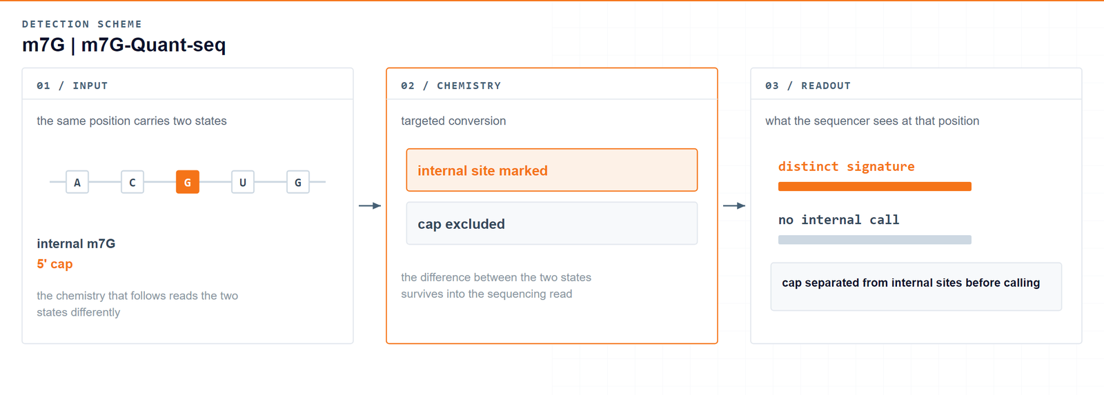
Internal m7G becomes measurable once the cap is taken out of the same signal.m7G-quant-seq: quantitative detection of RNA internal N7-methylguanosine, ACS Chemical Biology 2022;17(12) · DOI 10.1021/acschembio.2c00792
04Workflow
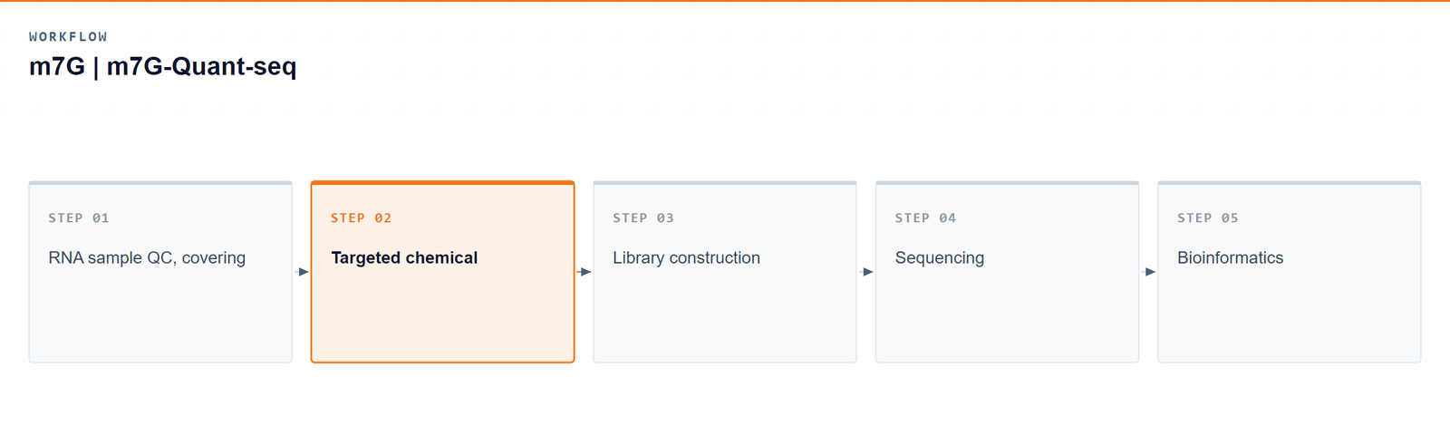
Five steps take a sample from RNA to a single-base internal m7G table:
- RNA sample QC, covering integrity and purity.
- Targeted chemical conversion of the RNA.
- Library construction from the converted material.
- Sequencing of the m7G-Quant-seq library.
- Bioinformatics: signature calling, internal-site annotation and stoichiometry estimation.
05Bioinformatics and deliverables
Reads are aligned to the reference transcriptome, m7G-Quant-seq conversion signatures are recognized, and internal m7G stoichiometry is estimated per site. The analysis set covers motif characterisation, transcript distribution, differential modification, functional enrichment and genome-browser visualization.
Deliverables
- Raw m7G mRNA sequencing data files (FASTQ), formatted for GEO/SRA submission.
- Sample and sequencing QC reports, sorted BAM alignment files and core mapping statistics.
- High-confidence single-base internal m7G site tables (XLSX) with coordinates, host gene and stoichiometry.
- Single-base m7G transcript feature distribution figures and consensus motif logos (PDF/PNG).
- Single-base differential m7G tables (XLSX) with fold change and significance.
- GO enrichment reports for m7G-modified genes (HTML/PDF/PNG).
- Genome-browser track files (bigWig/bedGraph) and a structured project report.
06Research applications
- Internal versus cap: separates the internal m7G population from the cap so the two are not reported as one number.
- Transcriptome-wide mapping: resolves internal m7G sites across protein-coding transcripts, and across lncRNA when that scope is selected.
- Translational control: connects a resolved internal site to mRNA translation and stability.
- Coding-region function: localises sites within CDS, UTR and splice-adjacent regions.
- Disease-oriented studies: identifies differentially modified sites as candidate biomarkers.
07Sample requirements
Storage. Cells and tissues: preserve in TRIzol or an RNA stabilisation solution, snap freeze in liquid nitrogen and store at −80 °C. RNA: resuspend in ethanol or RNase-free ultrapure water, store at −80 °C and avoid repeated freeze–thaw cycles.
Shipping. Place the sample in a 1.5 mL RNase-free microcentrifuge tube, seal it with parafilm or a cap lock, and ship on dry ice with adequate insulation.
| Sample type | Notes |
|---|---|
| Whole blood | EDTA tubes only; heparin is not compatible with the downstream procedure. |
| Cultured cells | Cell pellets are preferred. |
| Tissue | Fresh or frozen specimens; avoid necrotic material. |
| Total RNA | OD 260/280 ≥ 1.8 and RIN ≥ 7, with no visible degradation. Submit at least 10 µg total RNA. |
08FAQ
How is internal m7G told apart from the cap?
The conversion is designed to treat the two populations differently, so the internal sites carry a signature that the cap does not. That is what makes an internal-only site list possible.
What comes with each called site?
A genomic coordinate, the host gene, the functional region and a modification ratio, all mapped to the reference transcriptome.
Does the workflow use an antibody?
No. Detection rests on the targeted chemical conversion, so no antibody binding preference shapes which sites are recovered.
Why not use a bulk m7G measurement?
A bulk level cannot say which transcripts or which regions changed. Base-resolution calling is what supports a comparison between genes, conditions or regions within a transcript.
Can lncRNA be included in the same run?
Yes. Two scopes are available, mRNA alone or mRNA together with long non-coding RNA, analyzed in one experiment rather than as two separate projects.
09References
- m7G-quant-seq: quantitative detection of RNA internal N7-methylguanosine. ACS Chemical Biology 2022;17(12). DOI: 10.1021/acschembio.2c00792 PMID: 36398936.