
RNA m6A Modification Profile (MeRIP-Seq)
Antibody-based enrichment of m6A-carrying fragments, read as a transcript-level profile: a practical first pass when the question is which transcripts differ, not which nucleotide carries the mark.
01What the service reports
RNA m6A Modification Profile (MeRIP-Seq) is an antibody-based enrichment service. Fragments carrying m6A are pulled down with an anti-m6A antibody and sequenced, and the result is a transcript-level profile of where m6A is enriched.
The readout is a region, not a nucleotide. Enriched fragments map back to stretches of a transcript, and the profile describes how that enrichment shifts between samples. Because the signal comes from an antibody pull-down, the visible sites also carry the binding preference of the antibody.
That trade is the reason the method is still widely used: it scales across many samples and conditions at modest input, which makes it a natural screening step. Regions that look interesting in the profile are normally followed up with a single-base method. Arraystar delivers the full workflow, from RNA QC to the analyzed profile and comparison tables.
02Benefits
- Works across many samples: enrichment-based profiling is a practical way to compare m6A status over conditions, time points or cohorts.
- Transcript-level view: the profile shows which transcripts and regions carry enriched m6A, which is often the first question in a study.
- Well-established route: the antibody enrichment format is long established, so the analysis outputs and their interpretation are familiar.
- Pairs with single-base work: enrichment narrows the candidates and a single-base method resolves the individual sites inside them.
- Complete analysis: mapping, peak calling, annotation, differential comparison and visualization are part of the service.
| Service Name | RNA Class | Price |
|---|---|---|
| RNA m6A Modification Profile (MeRIP-Seq) | mRNA | |
| RNA m6A Modification Profile (MeRIP-Seq) | mRNA and lncRNA |
03Background and detection principle
MeRIP-Seq starts by fragmenting the RNA and incubating it with an antibody that recognises m6A. Antibody-bound fragments are captured, and the captured pool is sequenced alongside an input control. Comparing the two libraries shows which fragments were enriched, and from that comparison the profile is built.
Two boundaries follow from the chemistry and belong in the interpretation. First, an enriched fragment spans tens of nucleotides, so the profile localises enrichment to a region rather than resolving a position. Second, capture depends on antibody binding, so an m6A that is poorly recognized can be under-represented in the profile. Read with those limits in mind, the profile is a sound map of where to look next.
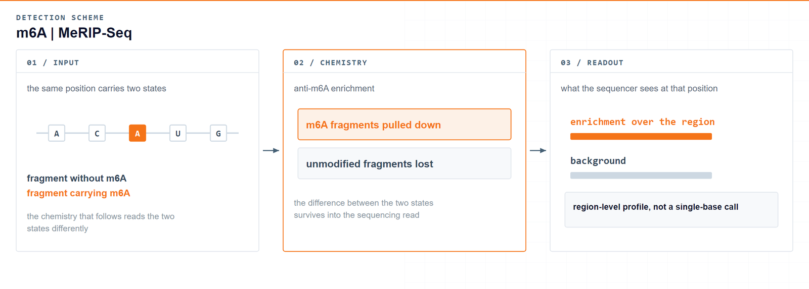
The antibody decides what is seen: MeRIP-Seq maps m6A to regions of a transcript and leaves position-level questions to the single-base methods.Meyer KD. et al., Cell 2012 · DOI 10.1016/j.cell.2012.05.003
04Workflow
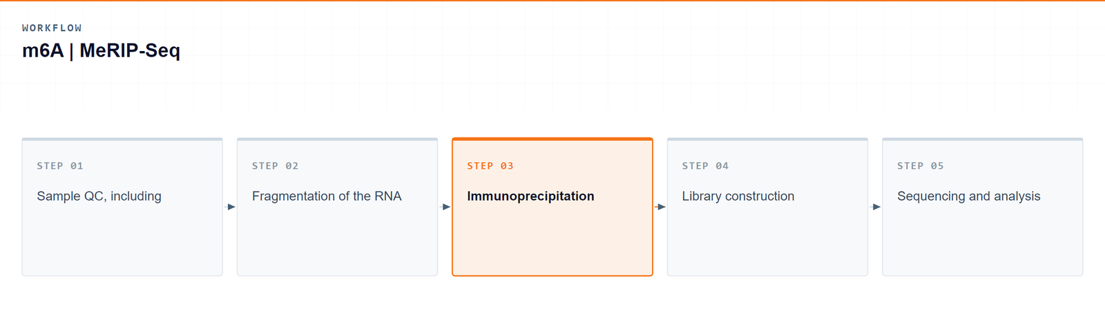
Five steps carry a sample from RNA to a transcript-level m6A profile:
- Sample QC, including integrity and purity checks.
- Fragmentation of the RNA into a size range suitable for capture.
- Immunoprecipitation with an anti-m6A antibody, against an input control.
- Library construction from the enriched and input fractions.
- Sequencing and analysis to call enriched regions and compare samples.
05Bioinformatics and deliverables
Reads are mapped and enriched regions are called against the input control, then annotated to transcript features. The analysis set covers peak annotation, motif analysis, distribution over transcript regions, differential enrichment between groups and genome-browser visualization.
Deliverables
- Raw m6A mRNA enrichment sequencing data files (FASTQ), formatted for GEO/SRA submission.
- QC reports and sorted, indexed BAM alignment files with core mapping statistics.
- m6A enrichment peak tables (XLSX) with genomic coordinates and annotated host genes.
- Peak distribution and consensus motif plots (PDF/PNG).
- Differential m6A enrichment tables (XLSX) between sample groups.
- GO enrichment reports for genes carrying enriched peaks (HTML/PDF/PNG).
- Genome-browser signal tracks (bigWig/bedGraph) and a structured project report.
06Research applications
- Cohort and clinical studies: comparing m6A enrichment across many patient or treatment samples is the classic use of this format.
- Cancer research: tumour and normal samples can be compared at the transcript level to identify genes whose m6A status changes.
- Development and differentiation: enrichment profiles across stages show which transcripts gain or lose m6A as cells commit.
- Stress and stimulus response: treated and untreated samples can be compared to see which regions respond at the m6A level.
- Screening before site-level work: the profile is used to choose the transcripts and regions that deserve single-base follow-up.
07Sample requirements
Storage. Cells and tissues: preserve in TRIzol or an RNA stabilisation solution, snap freeze in liquid nitrogen and store at −80 °C. RNA: resuspend in ethanol or RNase-free ultrapure water, store at −80 °C and avoid repeated freeze–thaw cycles.
Shipping. Place the sample in an RNase-free microcentrifuge tube, seal it against leakage, and ship on dry ice with adequate insulation.
| Sample type | Notes |
|---|---|
| Whole blood | EDTA tubes only; heparin is not compatible with the downstream procedure. |
| Cultured cells | Cell pellets are preferred. |
| Tissue | Fresh or frozen specimens; avoid necrotic material. |
| Total RNA | OD 260/280 ≥ 1.8 and RIN ≥ 7, with no visible degradation. Submit more than 10 µg total RNA (around 50 µg gives the best result; 10 µg is the practical lower limit). |
08FAQ
What if the study needs the position rather than the profile?
Then the work moves to single-base resolution. Alongside this profile, Arraystar provides m6A sequencing at single-base resolution (CAM-seq, GLORI2-seq), which reports where each site sits and what fraction of the RNA carries it, so transcripts flagged here can be resolved position by position on matched RNA.
Can MeRIP-Seq tell me the exact m6A position?
No. Enriched fragments cover a region, so the result is a profile of enrichment rather than a single resolved site. If the exact position and level are the conclusion, a single-base method is the right choice.
What role does the antibody play in the result?
Capture depends on the antibody binding an m6A in the fragment. An epitope that the antibody recognises poorly can be under-represented, so the visible profile carries the antibody’s binding preference as well as the underlying biology.
Should an input control be included?
Yes. Comparing the enriched fraction against an input control is what turns the sequencing data into enrichment calls, and it is part of the standard workflow.
How do I decide between MeRIP-Seq and a single-base method?
If the study asks which transcripts differ across many samples, enrichment is the efficient first pass. If the study asks where exactly the mark sits, start with a single-base method or follow the profile with one.
Which samples are accepted?
Total RNA, cultured cells, tissue or blood. Preparation notes and quality thresholds are listed on this page.
09References
- Meyer KD, Saletore Y, Zumbo P, Elemento O, Mason CE, Jaffrey SR. Comprehensive analysis of mRNA methylation reveals enrichment in 3′ UTRs and near stop codons. Cell 2012;149:1635–1646. DOI: 10.1016/j.cell.2012.05.003
- Dominissini D, Moshitch-Moshkovitz S, Schwartz S, et al. Topology of the human and mouse m6A RNA methylomes revealed by m6A-seq. Nature 2012;485:201–206. DOI: 10.1038/nature11112