
Single-Base RNA Modification Sequencing

Related Products
Related Services
Related Reviews

Single-Nucleotide RNA hm5C Modification Seq(WO-Seq)

Single-Nucleotide RNA hm5C Sequencing (WO-Seq) reads 5-hydroxymethylcytidine at single-nucleotide resolution without bisulfite. Peroxotungstate oxidizes hm5C to trihydroxylated thymine (thT); TGIRT reads thT as T during cDNA synthesis, so the hydroxymethylated cytosine appears as a C-to-T change at its own position.

Arraystar runs the route end to end: RNA QC, peroxotungstate oxidation, TGIRT reverse transcription and library construction, sequencing, and bioinformatics. Each reported site carries its host transcript, transcript region and a relative hm5C signal derived from the C-to-T fraction at that cytosine.

Benefits

In RNA, hm5C is a low-abundance mark that TET enzymes can produce by oxidizing m5C, so a readout that resolves the modified base itself is informative.

Single-base resolution: the C-to-T change sits at the hydroxymethylated cytosine, not at the center of an enriched interval.

Site-level quantification: the C-to-T signal at each cytosine is reported as a relative hm5C-associated value; a partly modified site keeps an intermediate value.

Bisulfite-free chemistry: the oxidation replaces the acid and alkali cycling of bisulfite conversion.

Antibody-free chemistry: peroxotungstate converts hm5C to thT while m5C and unmodified cytosine are largely retained. This avoids antibody-dependent enrichment and its binding bias; other sources of bias remain.

Functional annotation: every site is placed on its host transcript and transcript region.

Application-ready: supports TET biology, translation studies, development and disease models, and cross-method comparison.

One-stop service: sample QC, oxidation, library construction, sequencing and analysis inside one project, with professional bioinformatics support.

Service NameRNA ClassPrice
Single-Nucleotide RNA hm5C Sequencing (WO-Seq)mRNA
Single-Nucleotide RNA hm5C Sequencing (WO-Seq)mRNA & lncRNA

Background

5-Hydroxymethylcytidine (hm5C) is the oxidized form of m5C. TET enzymes have been shown to oxidize m5C to hm5C in RNA. RNA hm5C has been studied in relation to translation, neuronal biology and development; its distribution and functions remain under investigation, and it is present at low levels in mammalian samples.

Conventional bisulfite sequencing does not reliably distinguish hm5C from m5C: both can resist deamination and are read as C. The treatment also relies on acid and alkali cycling that damages RNA, and incomplete conversion in double-stranded regions can create false positives.

WO-Seq takes a different route. Peroxotungstate selectively oxidizes hm5C to trihydroxylated thymine (thT). The reaction works on single-stranded nucleic acid and is inefficient on double-stranded DNA, but RNA is not uniformly single-stranded; secondary and tertiary structure can reduce oxidation efficiency at some positions. TGIRT reads thT as T during cDNA synthesis, so the modified cytosine leaves the expected C-to-T signature. Under the reaction conditions, m5C and unmodified cytosine are largely retained.

Oxidation and reverse transcription are not necessarily complete, and observed T reads also include background mismatches and sequencing errors. The reported value is therefore a relative hm5C-associated C-to-T signal, not an absolute modification fraction. It is read within a batch and between libraries prepared under one protocol; coverage at the position determines whether a call is made. Sensitivity also depends on RNA abundance, structural context and conversion efficiency.

Key concept: WO-Seq is a bisulfite-free method for detecting hm5C in RNA at single-nucleotide resolution: under validated reaction conditions, peroxotungstate oxidizes hm5C to thT, TGIRT reads thT as T, and the expected C-to-T change marks the hydroxymethylated cytosine.
WO-Seq hm5C detection scheme

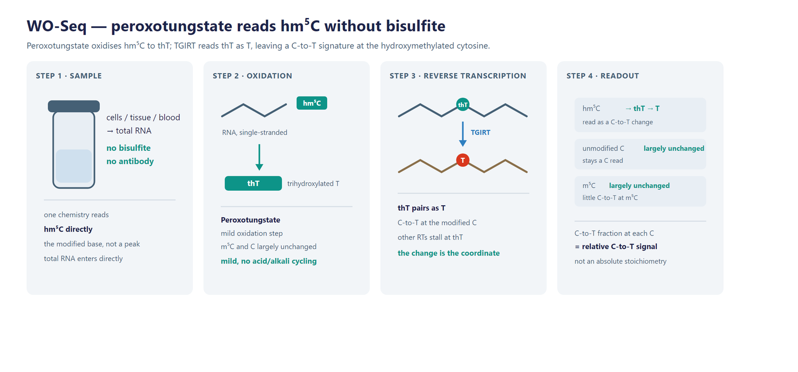
Figure 1. WO-Seq detection scheme. Peroxotungstate oxidizes hm5C to thT while m5C and unmodified cytosine are largely retained; TGIRT reads thT as T during cDNA synthesis, so the C-to-T change marks the hydroxymethylated position.

Workflow

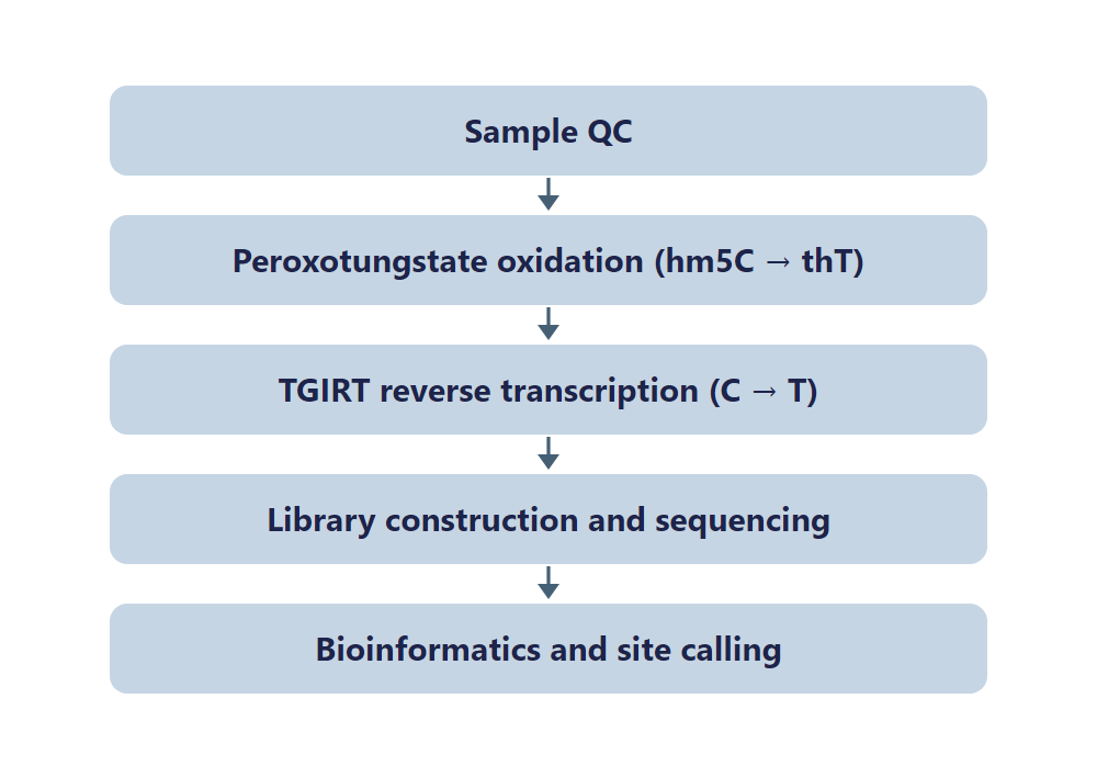
Five steps take a sample from RNA to a single-base hm5C table:

Sample QC: RNA integrity and purity are checked before the chemistry is started.

Oxidation: peroxotungstate converts hm5C to thT.

Reverse transcription: TGIRT reads thT as T, leaving a C-to-T signature at the modified cytosine.

Sequencing: the library is sequenced on an Illumina platform at a depth suited to position-level calling.

Bioinformatics: reads are aligned, every cytosine is scored for C-to-T support, and hm5C sites are annotated and quantified.
WO-Seq workflow

Figure 2. WO-Seq workflow, from sample QC through peroxotungstate oxidation and TGIRT reverse transcription to a single-base hm5C table.

Bioinformatics

Reads are aligned and every cytosine is scored for C-to-T support. Candidates are filtered by coverage and by local background; other cytosines on the same transcript are not automatically treated as unmodified, and the thresholds used are recorded in the project report. Between-group comparison accounts for coverage and background, and statistical testing requires biological replicates. The analysis adds the host transcript and transcript region of each site, plus genome-browser visualization of the C-to-T profile.

Interpretation stays within the design. A candidate is stronger when coverage is sufficient, the local background is clean and an independent assay supports it. Group comparisons need biological replicates. A change in T support can also come from RNA abundance, structure or conversion efficiency, so it is not by itself evidence of a change in hm5C. Report the design, controls and thresholds alongside the site list so the comparison can be read in context.

Deliverables

  • Raw FASTQ data for every library, formatted for GEO/SRA public database submission.
  • QC report with sorted BAM alignment files and core mapping statistics.
  • Single-base hm5C site table (XLSX) with coordinates, host transcript and relative hm5C-associated signal.
  • Per-site C-to-T support and coverage table (XLSX) for review of each call.
  • C-to-T profile and site distribution figures by transcript region (PDF/PNG).
  • Differential hm5C-associated signal table (XLSX) with fold change and significance.
  • Genome-browser track files (bigWig/bedGraph) and a structured project report.

Research Applications

  • TET biology: compare hm5C profiles after TET loss, gain or inhibition, and follow the mark downstream of m5C oxidation.
  • Translation: relate site-level hm5C to ribosome occupancy and to the translation of the transcripts that carry it.
  • Development: follow hm5C across differentiation and in models of neuronal development, where the mark has been studied.
  • Low-abundance marks: read hm5C without an antibody in samples where enrichment is difficult or background-prone.
  • Cancer and disease models: compare hm5C profiles between tumor and normal material or across disease subtypes.
  • Method comparison: set WO-Seq beside hMeRIP-seq or bisulfite-based maps generated from the same samples.

References

[1] Yuan F, Bi Y, Siejka-Zielinska P, et al. Bisulfite-free and base-resolution analysis of 5-methylcytidine and 5-hydroxymethylcytidine in RNA with peroxotungstate. Chemical Communications 2019;55:2328-2331. DOI: 10.1039/C9CC00274J. PMID: 30723849.

[2] Delatte B, Wang F, Ngoc LV, et al. Transcriptome-wide distribution and function of RNA hydroxymethylcytosine. Science 2016;351:282-285. DOI: 10.1126/science.aac5253. PMID: 26816380.

[3] Fu L, Guerrero CR, Zhong N, et al. Tet-mediated formation of 5-hydroxymethylcytosine in RNA. Journal of the American Chemical Society 2014;136:11582-11585. DOI: 10.1021/ja505305z. PMID: 25073028.

[4] Okamoto A, Sugizaki K, Nakamura A, et al. 5-Hydroxymethylcytosine-selective oxidation with peroxotungstate. Chemical Communications 2011;47:11231-11233. DOI: 10.1039/c1cc14782j. PMID: 21918786.

[5] Hayashi G, Koyama K, Shiota H, et al. Base-resolution analysis of 5-hydroxymethylcytosine by one-pot bisulfite-free chemical conversion with peroxotungstate. Journal of the American Chemical Society 2016;138:14178-14181. DOI: 10.1021/jacs.6b06428. PMID: 27766835.

Sample Requirements

Storage Guidelines

Cells and Tissues: Preserve in TRIzol or an RNA stabilization solution; snap freeze in liquid nitrogen and store at −80 °C.

RNA: Resuspend in ethanol or RNase-free ultrapure water; store at −80 °C and avoid multiple freeze-thaw cycles.

Shipping Instructions

Place the sample in a 1.5 mL RNase-free microcentrifuge tube.

Seal the tube with parafilm or a cap lock to ensure sample integrity.

Ship the package on dry ice with adequate insulation to maintain the required temperature.

Sample TypeNotes
Whole bloodUse EDTA tubes only, as heparin is not compatible with subsequent analytical procedures.
Cultured cellsSubmission of cell pellets is preferred to ensure high-quality material for processing.
TissueProvide fresh or frozen specimens, ensuring that necrotic material is strictly avoided.
Total RNAMaintain an OD 260/280 ratio ≥ 1.8 and RIN ≥ 7 with no visible degradation. Submit at least 10 µg total RNA.

FAQ

Why not read hm5C with bisulfite sequencing?

Conventional bisulfite sequencing does not reliably distinguish m5C from hm5C, because both can resist conversion and read as C. The treatment also uses acid and alkali cycling that damages RNA, and incomplete conversion in double-stranded regions can produce false positives. WO-Seq oxidizes hm5C directly, while m5C and unmodified cytosine are largely retained.

How does the chemistry distinguish hm5C from m5C?

Peroxotungstate uses hm5C as its substrate and converts it to thT; m5C is largely retained. TGIRT then reads thT as T, so the expected C-to-T change marks hm5C. At an m5C site the read typically stays C, which is also how an unmodified cytosine reads.

Does the assay give absolute hm5C levels?

No. Oxidation and reverse transcription are not necessarily complete, and T reads include background mismatches and sequencing errors. The value is a relative hm5C-associated C-to-T signal. It supports comparisons between samples processed under one protocol; an absolute fraction would need a calibrated method and a defined spike-in.

What determines whether a site is called?

Candidates are filtered by coverage and by local background; other cytosines on the same transcript are not automatically treated as unmodified. The thresholds used are recorded in the project report, and candidate sites can be checked independently. That record makes each call reviewable.

What research questions can a site-level hm5C map support?

It supports comparisons of hm5C positions after TET perturbation, between normal and disease samples, and alongside translation readouts. Because the signal is read at the cytosine, a study can follow one position rather than an enriched region.

What does a partly modified site look like?

It keeps an intermediate C-to-T signal. A site with more hm5C tends to give a higher signal, and a site modified on part of the pool sits between the background and the upper end of the assay’s range. The value is compared across samples, not converted to an absolute fraction.