
Single-Base tRNA Modification Sequencing

Related Products
Related Services
Related Reviews

Single-Nucleotide tRNA m3C Modification Seq(m3C-IP-seq)

m3C-IP-Seq is an antibody-based service that profiles m3C on tRNA. An m3C-specific antibody immunoprecipitates m3C-bearing fragments, then reverse-transcription misincorporation at the modified cytidine is resolved to a single base.

Arraystar Single-Nucleotide tRNA m3C Modification Seq(m3C-IP-Seq) is an end-to-end sample-to-data service for tRNA, from RNA sample QC and tRNA treatment through library construction, sequencing, and bioinformatics. Sites are localized within mature tRNA transcripts with relative coordinates, tRNA gene attribution, anticodon and isotype, and structural-domain localization.

Benefits

In tRNA, m3C influences structure and function, so site-level mapping is valuable. An antibody IP step enriches m3C-bearing fragments and a reverse-transcription readout registers each modified nucleotide at base resolution.

Single-base resolution: pinpoints m3C sites in tRNA.

Quantitative measurement: the misincorporation read-out yields reliable modification fractions.

Antibody-based enrichment: an m3C-specific antibody concentrates m3C-bearing fragments, so low-stoichiometry tRNA sites stand out from the unmodified background.

Functional annotation: Sites are localized within mature tRNA transcripts with relative coordinates, tRNA gene attribution, anticodon and isotype, and structural-domain localization.

Application-ready: supports decoding fidelity, codon use, aminoacylation, and tRNA structural biology across tRNA isotypes.

Service NamePrice
Single-Nucleotide tRNA m3C Modification Seq(m3C-IP-Seq)

Background

In tRNA, the modification stabilizes the folded L-shaped structure and fine-tunes anticodon–codon decoding, so base-resolution mapping within maturation loops and the anticodon is what distinguishes it. An m3C-specific antibody immunoprecipitates m3C-containing fragments, and reverse-transcription misincorporation at the modified cytidine is resolved to a single base, giving sensitive single-base m3C mapping. This antibody-based read-out maps each m3C site at single-base resolution.

The method was validated for tRNA, sites are localized within mature tRNA transcripts with relative coordinates, tRNA gene attribution, anticodon and isotype, and structural-domain localization. (Gao Y, Hou J, Wei S, et al. Nucleic Acids Research 2025;53(5)., DOI: 10.1093/nar/gkaf153).

Key concept: Single-Nucleotide tRNA m3C Modification Seq(m3C-IP-Seq) is a sequencing service in which an m3C-specific antibody immunoprecipitates m3C-containing fragments, and reverse-transcription misincorporation at the modified cytidine is called at single-base resolution.
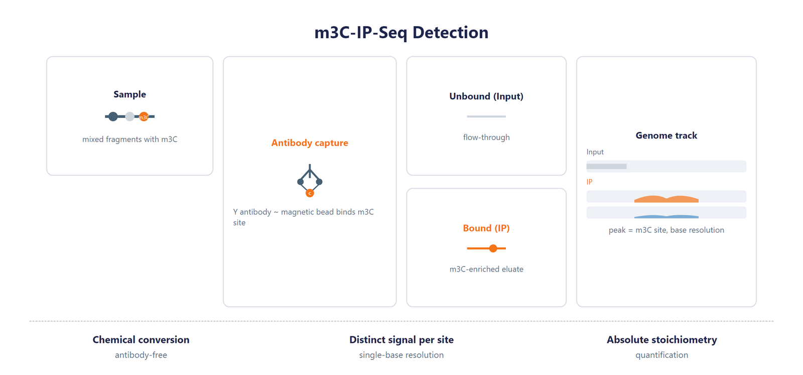
Single-Nucleotide tRNA m3C Modification Seq scheme

Figure 1. m3C-IP-Seq detection scheme. An antibody IP step enriches m3C-bearing fragments, and a reverse-transcription readout registers the modified nucleotide at base resolution.

Workflow

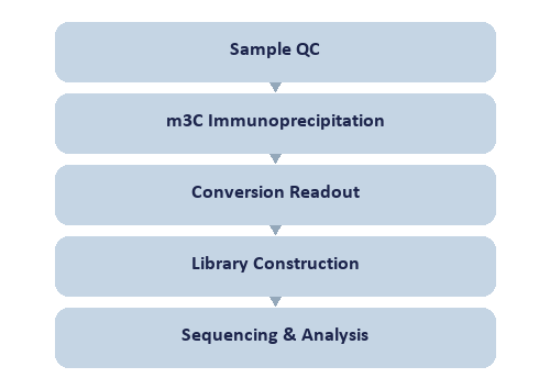
A streamlined workflow carries the sample to base-resolution N3-methylcytidine (m³C) datasets:
Single-Nucleotide tRNA m3C Modification Seq workflow

Figure 2. Single-Nucleotide tRNA m3C Modification Seq workflow.

Bioinformatics

Downstream analysis maps reads to a curated reference, calls m3C-IP-Seq misincorporation signatures, and reports m3C stoichiometry per position with motif, differential, enrichment, and genome-browser outputs.

Deliverables

  • Raw m³C tRNA sequencing FASTQ data files, formatted for GEO/SRA public database submission.
  • QC report with BAM alignment files and key mapping metrics.
  • High-confidence single-base m³C tRNA site tables (XLSX) with coordinates, anticodon, isotype, and stoichiometry.
  • Single-base m³C distribution plots across tRNA domains and consensus m³C motif logos (PDF/PNG).
  • Single-base resolution differential m³C tRNA site tables (XLSX) with fold change and significance.
  • Genome browser-compatible m³C signal track files (bigWig/bedGraph) and a structured tRNA report.

Research Applications

  • tRNA decoding fidelity: maps the modification within anticodon and wobble regions.
  • Codon use & aminoacylation: relates base-resolution modification to charging and translation.
  • tRNA structural biology: localizes sites within D-loop, anticodon, TΨC, and acceptor stem.
  • Isotype-resolved analysis: compares modification across tRNA isotypes and isoacceptors.
  • Disease-oriented studies: finds differentially modified tRNA sites as biomarkers.

References

[1] Gao Y, Hou J, Wei S, et al. Transcriptome-wide mapping of N3-methylcytidine modification at single-base resolution. Nucleic Acids Research 2025;53(5). DOI: 10.1093/nar/gkaf153.

Sample Requirements

Sample Storage

For cells/tissue: use TRIzol or an RNA-stabilizing reagent, quick-freeze in liquid nitrogen, and keep at –80 °C.

For RNA: dissolve in ethanol or RNase-free water, store at –80 °C, and limit freeze–thaw cycles.

Dispatch Guidelines

Transfer each sample into a 1.5 mL nuclease-free tube.

Close the tube securely with parafilm or a cap lock to preserve integrity.

Send on dry ice with sufficient insulation to sustain the required temperature.

Sample TypeNotes
Whole bloodUse EDTA tubes only, as heparin is not compatible with subsequent analytical procedures.
Cultured cellsSubmission of cell pellets is preferred to ensure high-quality material for processing.
TissueProvide fresh or frozen specimens, ensuring that necrotic material is strictly avoided.
Total RNAMaintain an OD 260/280 ratio ≥ 1.8 and RIN ≥ 7 with no visible degradation. Submit at least 200 µg total RNA.

FAQ

Which tRNA features are annotated?

Sites are localized within mature tRNA transcripts with relative coordinates, tRNA gene attribution, anticodon and isotype, and structural-domain localization. This lets you interpret each site in the context of its host tRNA isotype and structural domain. The method supports both mRNA and tRNA workflows, so you can choose the readout that matches your study.

Does this method measure the modification at single-base resolution?

Yes. An m3C-specific antibody immunoprecipitates m3C-containing fragments, and reverse-transcription misincorporation at the modified cytidine is resolved to a single base, giving sensitive single-base m3C mapping. This provides single-base resolution and absolute stoichiometry for each site in tRNA. An m3C-specific antibody drives the enrichment, and reverse-transcription misincorporation provides the single-base call, so the reported position and stoichiometry stay reliable for downstream functional analyses.

How does this method compare with antibody-free tRNA profiling?

Antibody-free chemical and signature assays read the modification directly, while antibody enrichment adds sensitivity for low-stoichiometry m3C positions. m3C-IP-Seq puts an m3C-specific antibody in front of the reverse-transcription read-out, so the same position is called at single-base resolution and with per-site stoichiometry. The two routes are complementary, and running m3C-IP-Seq alongside a cleavage-based m3C assay gives orthogonal confirmation for sites that are being reported for the first time.

How does this service support research on m3C modification?

By profiling m3C with high accuracy, the service helps researchers explore its biological significance, offering insight for studying RNA function and regulation.

Why is accurate m3C detection a valuable research asset?

Reliable m3C profiling strengthens research findings and supports advancement of understanding in RNA biology.