
Arraystar GlycoRNA Blotting Service uses lectin-based detection (LBD) to detect endogenous glycoRNAs following RNA separation and membrane transfer. By exploiting the natural glycan-binding specificity of lectins, the assay provides a simple and rapid approach for comparing glycoRNA abundance and molecular-size patterns across biological samples.
The service is applicable to cultured cells, tissues, and biofluids, providing an experimental tool for investigating glycoRNA biology and comparing glycoRNA-associated signals across biological conditions.
Benefits
Native-state detection: Detect endogenous glycoRNAs without artificial metabolic labeling such as Ac₄ManNAz.
Lectin-based glycan recognition: Uses lectin binding to detect glycan-containing RNA species.
Simple and rapid workflow: Avoids prolonged metabolic incubation and complex labeling procedures.
Broad sample compatibility: Suitable for cultured cells, tissues, and biofluids.
Comparative glycoRNA profiling: Enables comparison of glycoRNA-associated signals across experimental groups and biological samples.
Practical validation tool: Provides an orthogonal biochemical approach for glycoRNA research and exploratory validation.
| Service Name | Price |
|---|---|
| Arraystar GlycoRNA Blotting Service |
Background
Arraystar GlycoRNA Blotting Service uses the natural glycan-binding specificity of lectins to detect glycoRNA-associated signals on Northern blots.
The assay follows a streamlined workflow:
RNA extraction → gel electrophoresis → membrane transfer → lectin probing → signal detection
Unlike metabolic labeling approaches, the lectin-based strategy does not require artificial metabolic incorporation such as Ac₄ManNAz. This enables detection of endogenous glycoRNAs without prolonged metabolic incubation.
Why GlycoRNA Blotting?
The method provides a practical approach for researchers who want to:
- Compare glycoRNA abundance between biological conditions
- Examine glycoRNA patterns across tissues or cell types
- Investigate glycoRNA-associated changes in disease models
- Explore glycoRNA signals in biofluids
- Generate biochemical evidence for glycoRNA-related studies
Key concept: GlycoRNA blotting detects endogenous glycoRNAs in their native state by probing a Northern blot with lectins, whose natural glycan-binding specificity recognizes the glycan moiety rather than the RNA sequence, so no artificial metabolic labeling such as Ac₄ManNAz is required.
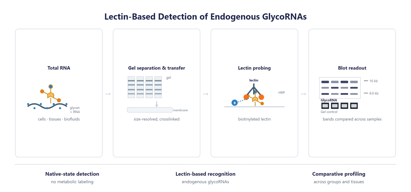
Figure 1. Principle of GlycoRNA lectin-based detection. Total RNA is separated by gel electrophoresis, transferred to a membrane, crosslinked, and probed with a lectin-based detection system to visualize glycoRNA-associated signals.
Workflow
Arraystar GlycoRNA Blotting Service provides an integrated experimental workflow from RNA preparation through glycoRNA detection and result presentation.
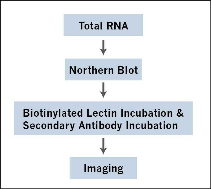
Figure 2. GlycoRNA Blot workflow. Total RNA extracted from cells or tissues is separated by gel electrophoresis, transferred onto a membrane, crosslinked, and incubated with a lectin primary probe followed by a secondary detection system to visualize glycoRNA bands.
1. RNA Sample Preparation: Total RNA is extracted from the submitted biological samples or supplied as purified RNA.
2. RNA Gel Electrophoresis: RNA samples are separated by gel electrophoresis to resolve RNA species according to molecular size.
3. Membrane Transfer: Separated RNA is transferred onto a membrane for subsequent detection and crosslinked to the membrane.
4. Lectin Probing: A lectin probe is applied to the membrane to recognize glycan-associated RNA species.
5. Signal Detection: A secondary detection system is used to visualize lectin-associated signals and generate glycoRNA blot images.
6. Comparative Analysis: GlycoRNA-associated band patterns and signal intensities can be compared across samples to assess differences in glycoRNA abundance and molecular-size distribution.
Bioinformatics
GlycoRNA blotting provides a direct visual readout of lectin-detectable glycoRNA-associated signals. Results can be used to compare signal patterns between biological samples and experimental conditions.
Deliverables
- Sample QC report with RNA integrity and concentration metrics for all submitted samples.
- GlycoRNA blot images with molecular-size markers and gel loading controls.
- Comparative signal summary tables across samples, conditions, and experimental groups.
- Project summary report covering the workflow, results, and interpretation notes.
Demo results
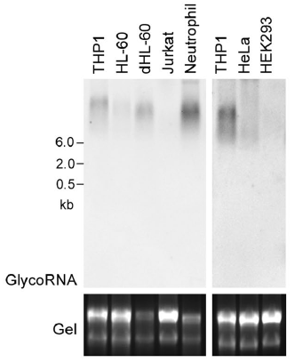
Demo 1. Images of detected glycoRNAs in human cell lines.
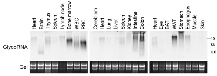
Demo 2. Images of detected glycoRNAs in mouse tissues.
Research Applications
Arraystar GlycoRNA Blotting Service can support a broad range of studies investigating RNA glycosylation and glycoRNA biology.
Cell-Based GlycoRNA Studies
Compare glycoRNA-associated signals between cultured cell populations under different experimental conditions.
Potential applications:
- Treatment-response studies
- Cellular phenotype-associated glycoRNA changes
- Comparison of control and genetically modified cells
- Exploratory glycoRNA profiling
Tissue-Specific GlycoRNA Profiling
Compare glycoRNA-associated signals among different tissues to investigate tissue-specific glycoRNA patterns.
The service has demonstrated glycoRNA detection in mouse tissues.
Disease-Associated GlycoRNA Research
Compare glycoRNA-associated signals between healthy and disease-associated biological samples to identify changes potentially associated with disease biology.
Biofluid GlycoRNA Studies
The assay is compatible with biofluids, enabling exploratory investigation of glycoRNA-associated signals in extracellular biological samples.
GlycoRNA Validation
GlycoRNA blotting can provide an independent biochemical readout to complement other glycoRNA detection or profiling strategies.
Recommended Experimental Strategy
For discovery studies, researchers may combine:
GlycoRNA blotting → glycoRNA profiling/sequencing → candidate validation
This complementary strategy can provide both comparative biochemical evidence and molecular-level characterization.
Sample Requirements
Storage Guidelines
Cells and Tissues: Preserve in TRIzol or an RNA stabilization solution; snap freeze in liquid nitrogen and store at −80 °C.
RNA: Resuspend in ethanol or RNase-free ultrapure water; store at −80 °C and avoid multiple freeze-thaw cycles.
Use high-quality RNA with minimal degradation, and provide sufficient material for the planned number of experimental conditions and replicates.
Shipping Instructions
Place the sample in a 1.5 mL RNase-free microcentrifuge tube.
Seal the tube with parafilm or a cap lock to ensure sample integrity.
Ship the package on dry ice with adequate insulation to maintain the required temperature.
| Sample Type | Notes |
|---|---|
| Whole blood / biofluid | Use EDTA tubes only, as heparin is not compatible with subsequent analytical procedures; plasma and serum are also accepted. |
| Cultured cells | Submission of cell pellets is preferred to ensure high-quality material for processing. |
| Tissue | Provide fresh or frozen specimens, ensuring that necrotic material is strictly avoided. |
| Total RNA | Maintain an OD 260/280 ratio ≥ 1.8 and RIN ≥ 7 with no visible degradation. Submit at least 10 µg total RNA. |
Because sample requirements may vary with sample type, RNA abundance, and experimental design, please contact Arraystar before submission for project-specific recommendations.
FAQ
What is glycoRNA?
GlycoRNAs are RNA molecules associated with glycans. GlycoRNA research investigates the molecular characteristics and biological functions of these glycan-associated RNA species, together with how their abundance and size distribution change across cell types, tissues, and experimental conditions. Endogenous glycoRNAs can be detected directly in total RNA, so their levels can be compared between biological samples without introducing artificial labels.
How are glycoRNAs detected by blotting?
In the Arraystar GlycoRNA Blotting Service, RNA is separated by gel electrophoresis, transferred to a membrane, crosslinked, and probed with a lectin-based detection system to visualize glycoRNA-associated signals. Lectins bind the glycan moiety of the RNA, so the resulting blot reports glycoRNA abundance and molecular-size patterns across samples.
Does GlycoRNA blotting require metabolic labeling?
No. The lectin-based detection approach does not require artificial metabolic labeling such as Ac₄ManNAz. Because lectins recognize glycans naturally, endogenous glycoRNAs can be detected directly in the submitted samples, avoiding prolonged metabolic incubation and the labeling steps that accompany it.
What is the advantage of lectin-based glycoRNA detection?
Lectins provide natural glycan-binding specificity, enabling detection of glycan-associated RNA without requiring metabolic incorporation of artificial sugar analogs. This keeps the workflow simple and rapid, works with cells, tissues, and biofluids, and provides an orthogonal biochemical readout that can complement sequencing-based glycoRNA profiling and validation studies.
What sample types are compatible with GlycoRNA Blotting?
The service can be applied to cultured cells, tissues, and biofluids. Purified RNA can also be submitted for projects in which RNA extraction has already been completed. Total RNA samples should show no visible degradation, and sufficient material should be provided for the planned number of conditions and replicates.
Can GlycoRNA blotting identify individual glycoRNA transcripts?
GlycoRNA blotting primarily provides a biochemical detection and comparative profiling readout rather than transcript-level identification. Individual glycoRNA species generally require complementary molecular characterization approaches, so blotting is best used together with profiling or sequencing methods when transcript-level information is needed.
References
[1] Flynn RA, Pedram K, Malaker SA, et al. Small RNAs are modified with N-glycans and displayed on the surface of living cells. Cell 2021;184(12):3109-3124.e22. DOI: 10.1016/j.cell.2021.04.023.
[2] Xie Y, Chai P, Till NA, et al. The modified RNA base acp³U is an attachment site for N-glycans in glycoRNA. Cell 2024;187(19):5228-5237.e12. DOI: 10.1016/j.cell.2024.07.044.
[3] Li Y, Qian Y, Li X, et al. Lectin-based detection and expression profiling of native glycoRNAs. Scientific Reports 2026;16(1). DOI: 10.1038/s41598-026-40291-2.
[4] Yi L, Zhou Y, Zhang C, et al. GlycoRNA research: from unknown unknowns to known unknowns. Protein & Cell 2026;17(2):1-20. DOI: 10.1093/procel/pwaf102.