
Single-Nucleotide rRNA 2′-O-Methylation Modification Seq(RiboMeth-Seq) maps 2′-O-methylation (Nm) across ribosomal RNA at single-nucleotide resolution. Alkaline treatment fragments the RNA at random positions; a 2′-O-methylated ribose blocks cleavage of the neighbouring phosphodiester bond, so an Nm position appears as a local gap in the fragment-end profile.
Arraystar runs the whole route as one sample-to-data workflow, from RNA QC and random fragmentation through library construction, sequencing and bioinformatics, and returns the rRNA Nm landscape as per-site tables on 18S, 28S and 5.8S coordinates.
Benefits
On rRNA, 2′-O-methylation is written at defined positions by the guide snoRNA system, so a per-site map is informative.
Single-base resolution: pinpoints each Nm position on 18S, 28S and 5.8S rRNA.
Quantitative stoichiometry: the depth of the gap at a position, read from its 5′/3′ coverage profile, is reported as its methylation ratio, so partially modified sites keep their intermediate value.
Antibody-free chemistry: the call follows alkaline cleavage of unmodified ribose, so no antibody enrichment enters the readout.
Functional annotation: sites are reported on standard rRNA coordinates with their rRNA region and structural context.
Application-ready: supports studies of ribosome heterogeneity, translation control, development and cancer.
One-stop service: sample QC, fragmentation, library construction, sequencing and analysis inside one project, with professional bioinformatics support.
| Service Name | Price |
|---|---|
| Single-Nucleotide rRNA 2′-O-Methylation Modification Seq(RiboMeth-Seq) |
Background
2′-O-methylation (Nm) is a methylation at the 2′-hydroxyl of the ribose. On ribosomal RNA it is written by fibrillarin together with box C/D guide snoRNAs, each of which selects its target position by base pairing, and it concentrates at positions that shape ribosome assembly and translation.
A 2′-O-methylated ribose makes the phosphodiester bonds next to it resistant to alkaline hydrolysis. RiboMeth-seq turns that difference into a readout: RNA is fragmented at random, the fragments that survived are sequenced, and the 5′/3′ fragment ends are counted position by position along the rRNA. fragment ends sit at every unmodified ribose, and an Nm position appears as a local gap in that profile, and the depth of the gap gives its methylation level.
Key concept: Single-Nucleotide rRNA 2′-O-Methylation Modification Seq(RiboMeth-Seq) is a chemical sequencing method in which alkaline random fragmentation is blocked by a 2′-O-methylated ribose, so the cleavage-resistant positions of rRNA report Nm sites at single-nucleotide resolution.
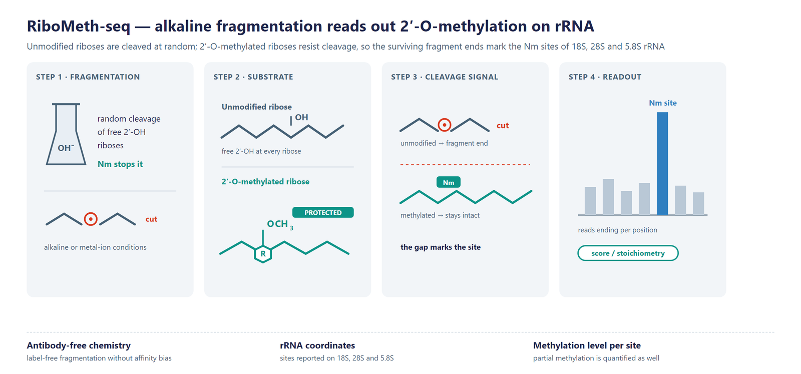
Figure 1. RiboMeth-seq detection scheme on rRNA: alkaline random fragmentation cuts unmodified positions, a 2′-O-methylated ribose blocks cleavage, and an Nm position appears as a local gap in the fragment-end profile.
Workflow
Five steps from RNA sample to single-base rRNA 2′-O-methylation data:
Sample QC: assess RNA integrity, purity and visible degradation before fragmentation.
Random fragmentation: treat the RNA under alkaline conditions so unmodified positions are cut and methylated positions are held.
Library construction: convert the surviving fragments into a sequencing library.
Sequencing: sequence on an Illumina platform to a depth suited to rRNA Nm calling.
Bioinformatics: align reads to the rRNA reference, score every position and annotate the sites that carry Nm.
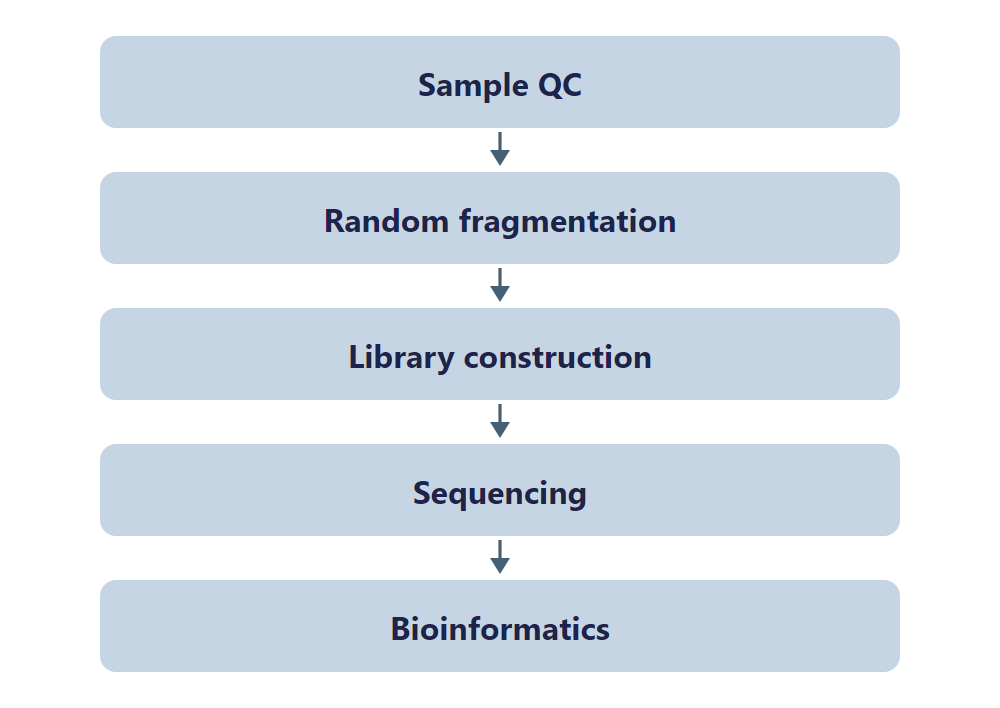
Figure 2. RiboMeth-seq workflow, from RNA sample through alkaline fragmentation and sequencing to single-base rRNA Nm data.
Bioinformatics
The pipeline aligns reads to the rRNA reference, counts fragment ends at every position and converts the surviving fraction into a methylation level for that site. Analysis adds the structural context of each position, site-level comparison between groups and genome-browser visualization.
Deliverables
- Raw FASTQ data files, ready for GEO/SRA public database submission.
- QC report with BAM alignment files and core mapping statistics.
- Single-base rRNA 2′-O-methylation site tables (XLSX) with rRNA coordinates and methylation level.
- Nm distribution figures across 18S, 28S and 5.8S rRNA (PDF/PNG).
- Differential Nm modification tables (XLSX) with fold change and significance.
- Annotation tables linking each Nm position to its rRNA region.
- Genome-browser track files (bigWig/bedGraph) and a structured project report.
Research Applications
- Ribosome heterogeneity: compare Nm levels site by site to describe ribosomes of different composition.
- Translation control: relate site-specific methylation to how particular mRNA sets are translated.
- Signaling and oncogenes: follow how rRNA methylation responds to upstream signaling programs.
- Development and differentiation: track Nm dynamics as cells move between states and lineages.
- Cancer research: profile Nm across patient samples and test whether it tracks subtype, grade or treatment response.
- Enzyme and guide studies: read out how fibrillarin levels or guide-snoRNA changes shift methylation at individual sites.
References
[1] Profiling of ribose methylations in RNA by high-throughput sequencing. Angewandte Chemie International Edition 2015; DOI: 10.1002/anie.201408362.
[2] Illumina-based RiboMethSeq approach for mapping of 2′-O-Me residues in RNA. Nucleic Acids Research 2016; DOI: 10.1093/nar/gkw547.
[3] Identification of sites of 2′-O-methylation vulnerability in human ribosomal RNAs by systematic mapping. Scientific Reports 2017; DOI: 10.1038/s41598-017-09734-9.
Sample Requirements
Storage Guidelines
Cells and Tissues: Preserve in TRIzol or an RNA stabilization solution; snap freeze in liquid nitrogen and store at -80 °C.
RNA: Resuspend in ethanol or RNase-free ultrapure water; store at -80 C and avoid multiple freeze-thaw cycles.
Shipping Instructions
Place the sample in a 1.5 mL RNase-free microcentrifuge tube.
Seal the tube with parafilm or a cap lock to ensure sample integrity.
Ship the package on dry ice with adequate insulation to maintain the required temperature.
| Sample Type | Notes |
|---|---|
| Whole blood | Use EDTA tubes only, as heparin is not compatible with subsequent analytical procedures. |
| Cultured cells | Submission of cell pellets is preferred to ensure high-quality material for processing. |
| Tissue | Provide fresh or frozen specimens, ensuring that necrotic material is strictly avoided. |
| Total RNA | Maintain an OD 260/280 ratio ≥ 1.8 and RIN ≥ 7 with no visible degradation. Submit at least 10 µg total RNA. |
FAQ
What does 2′-O-methylation do on the ribosome?
Fibrillarin writes it onto rRNA together with box C/D guide snoRNAs, each guide selecting one position by base pairing. The methyl groups sit preferentially at the ribosome functional centers and at the interfaces between subunits, where they influence assembly, decoding accuracy and the efficiency of protein synthesis.
Why does alkaline fragmentation read rRNA Nm sites so directly?
Alkaline conditions cut the RNA backbone wherever the ribose carries a free 2′-hydroxyl. A 2′-O-methylated ribose has no such hydroxyl, so the bonds next to it survive, and an Nm position appears as a gap in the fragment-end profile, and the depth of the gap reports how often the site was methylated.
How does this differ from NM-seq, which also maps Nm?
Both methods read the same modification by different chemistry. RiboMeth-seq cleaves randomly under alkaline conditions and scores the positions that resist cleavage, which suits abundant, structured RNA such as rRNA. NM-seq instead enriches Nm fragments through periodate oxidation-elimination-dephosphorylation cycles and is aimed at transcript-level RNA such as mRNA and lncRNA.
Are all rRNA Nm sites methylated to the same level?
No. Across human cell lines the sites fall into groups: about a third are methylated at a stable level, roughly half vary only slightly, and the remainder are clearly variable. Stable positions tend to sit in the functional centers, variable ones at the periphery, which is how ribosomes of different composition arise.
Can several conditions or groups be compared in one project?
Yes. All libraries are analyzed against one rRNA coordinate system and one scoring threshold, so sites keep the same numbering across groups and the differential table reports fold change and significance for every position without any manual alignment of coordinates.
How is the methylation level at a site computed?
At each position the pipeline combines the 5′ and 3′ end counts with the local coverage, following the published MethScore-type scoring, and reports the result as a level between zero and one. The result is a level between zero and one, so a site methylated on only part of the rRNA molecules returns an intermediate value rather than a yes/no call.