
Single-Base tRNA Modification Sequencing

Related Products
Related Services
Related Reviews

Single-Nucleotide tRNA m7G&m3C Modification Seq(Alkaniline-seq)

Single-Nucleotide tRNA m7G&m3C Modification Seq (AlkAniline-Seq) reports m7G and m3C on tRNA at single-nucleotide resolution. Heat under alkaline conditions detaches the labile base, aniline cuts the chain at the abasic site that is left behind, and the 5′-phosphate created at the neighbouring residue becomes the starting point of every read.

Arraystar runs the route as one sample-to-data workflow, from RNA sample QC and the cleavage chemistry through library construction, sequencing and bioinformatics, and returns a per-site tRNA table in which each entry carries its position within the mature tRNA, the structural domain it belongs to and the tRNA it was read on.

Benefits

In tRNA, m7G and m3C sit in the loops that set folding and decoding, so a position-level map of each is informative.

Single-base resolution: pinpoints m7G and m3C positions on the mature tRNA.

Quantitative measurement: the fraction of reads starting immediately 3′ of the cut is reported as the site level, so a partially modified position keeps its intermediate value.

Two marks in the same assay: m7G and m3C are read together, so the two site lists can be compared directly.

No antibody dependence: the call follows the cleavage chemistry, and total RNA enters the reaction without a tRNA fractionation step.

Functional annotation: sites are localized within mature tRNA transcripts with relative coordinates, tRNA gene attribution, anticodon and isotype, and structural-domain localization.

Application-ready: supports decoding fidelity, codon use, aminoacylation, and tRNA structural biology across tRNA isotypes.

One-stop service: sample QC, cleavage chemistry, library construction, sequencing and analysis inside one project, with professional bioinformatics support.

Service NamePrice
Single-Nucleotide tRNA m7G Modification Seq (AlkAniline-Seq)

Background

tRNA carries both marks this assay reads. m7G sits in the variable loop of a defined subset of tRNAs, where the METTL1/WDR4 complex writes it, and m3C sits mostly in the anticodon loop (position 32), with additional sites in the variable arm (C47d) and the D-arm (C20); METTL6 and the METTL2A/2B enzymes write it on different tRNA families. Both methyl groups sit on a base whose bond to the ribose is unstable compared with an ordinary residue.

AlkAniline-Seq turns that instability into a position. Heat under alkaline conditions removes the modified base and leaves an abasic site; aniline drives beta-elimination there and cuts the chain; and pre-existing 5′-phosphates are removed by dephosphorylation, so the 5′-phosphate left on the residue next to the modified base is the only end in the reaction that accepts an adapter. Because the library is built outwards from each cut, a read starts exactly where the chemistry stopped, and the mark is identified by the parental base: guanosine for m7G, and a cytidine cut is annotated as m3C only at positions known to carry m3C, because ho⁵C shares the same labile bond.

Key concept: Single-Nucleotide tRNA m7G Modification Seq (AlkAniline-Seq) is a chemistry-based sequencing method in which the labile base of m7G or m3C is removed under heat and alkaline conditions, aniline cleaves the tRNA backbone at the resulting abasic site, and the 5′-phosphate created at the next residue directs adapter ligation, so the cut reports the modification position on the tRNA.
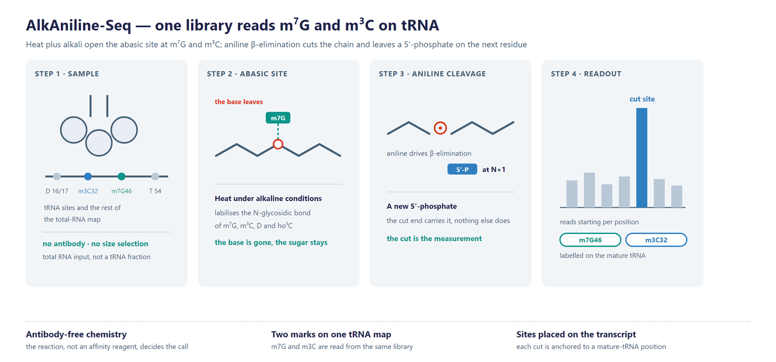
Single-Nucleotide tRNA m7G Modification Seq (AlkAniline-Seq) scheme

Figure 1. AlkAniline-Seq detection scheme on tRNA. Heat and alkali generate the abasic site at m7G and m3C, aniline cleavage leaves a 5′-phosphate at the neighbouring residue, and adapter ligation from that phosphate is what turns each cut into a readable tRNA position.

Workflow

Five steps take a sample from RNA to a single-base tRNA m7G and m3C table:

Sample QC: RNA integrity and purity are checked before the chemistry is started.

Abasic-site formation and dephosphorylation: the RNA is held under heat and alkaline conditions so the labile base leaves its ribose, after which pre-existing 5′-phosphates are removed.

Aniline cleavage and library construction: aniline cuts the chain at every abasic site and the resulting 5′-phosphate ends are ligated to sequencing adapters.

Sequencing: the library is sequenced on an Illumina platform at a depth suited to position-level calling on tRNA.

Bioinformatics: reads are aligned to a tRNA reference, cut positions are scored nucleotide by nucleotide, and m7G and m3C sites are annotated on the mature tRNA and quantified.
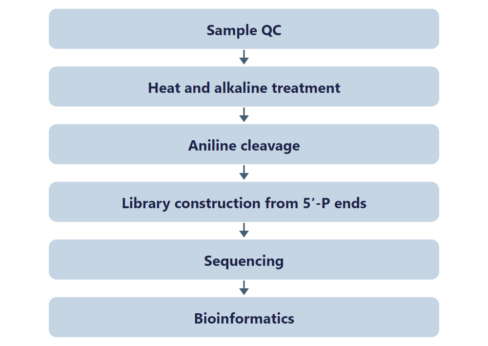
Single-Nucleotide tRNA m7G Modification Seq (AlkAniline-Seq) workflow

Figure 2. AlkAniline-Seq workflow, from sample QC through abasic-site formation, aniline cleavage and sequencing to a single-base tRNA m7G and m3C table.

Bioinformatics

Reads are aligned to a tRNA reference, cut positions are scored at every nucleotide, and guanosine positions are reported as m7G while cytidine positions are reported as m3C, so the two marks keep separate site lists. Analysis adds the tRNA identity and the structural domain of each position, site-level comparison between sample groups and genome-browser visualization.

Deliverables

  • Raw FASTQ data files, ready for GEO/SRA public database submission.
  • QC report with BAM alignment files and core mapping statistics.
  • Single-base tRNA m7G site tables (XLSX) with tRNA coordinates, tRNA identity and site level.
  • Single-base tRNA m3C site tables (XLSX) with tRNA coordinates, tRNA identity and site level.
  • Site distribution figures by tRNA structural domain and by isotype (PDF/PNG).
  • Differential modification tables (XLSX) with fold change and significance.
  • Genome-browser track files (bigWig/bedGraph) and a structured project report.

Research Applications

  • Translational control: test whether a change in m7G site level on a tRNA family is mirrored by ribosome occupancy and by the translation of the codons that family decodes.
  • m3C and codon-biased translation: follow m3C sites in the anticodon loop and relate them to serine codon-biased translation, cell cycle progression and the response to DNA-damaging treatment.
  • Writer-enzyme studies: read how loss or gain of METTL1/WDR4 shifts m7G, and how METTL6 or METTL2A/2B shifts m3C, on individual tRNA families.
  • Development and disease models: profile tRNA site levels in models of m7G-related disease and in cells that exit or re-enter the cell cycle.
  • Cancer research: compare tRNA site profiles between tumour and normal tissue, or between treatment-sensitive and resistant material.
  • Method cross-validation: set the chemical call beside antibody-based or cleavage-based tRNA maps generated from the same samples.

References

[1] AlkAniline-Seq: profiling of m7G and m3C RNA modifications at single nucleotide resolution. Angewandte Chemie International Edition 2018; DOI: 10.1002/anie.201810946.

[2] Mettl1/Wdr4-mediated m7G tRNA methylome is required for normal mRNA translation and embryonic stem cell self-renewal and differentiation. Molecular Cell 2018; DOI: 10.1016/j.molcel.2018.06.001.

[3] m3C32 tRNA modification controls serine codon-biased mRNA translation, cell cycle, and DNA-damage response. Nature Communications 2024; DOI: 10.1038/s41467-024-50161-y.

Sample Requirements

Storage Guidelines

Cells and Tissues: Preserve in TRIzol or an RNA stabilization solution; snap freeze in liquid nitrogen and store at -80 °C.

RNA: Resuspend in ethanol or RNase-free ultrapure water; store at -80 °C and avoid multiple freeze-thaw cycles.

Shipping Instructions

Place the sample in a 1.5 mL RNase-free microcentrifuge tube.

Seal the tube with parafilm or a cap lock to ensure sample integrity.

Ship the package on dry ice with adequate insulation to maintain the required temperature.

Sample TypeNotes
Whole bloodUse EDTA tubes only, as heparin is not compatible with subsequent analytical procedures.
Cultured cellsSubmission of cell pellets is preferred to ensure high-quality material for processing.
TissueProvide fresh or frozen specimens, ensuring that necrotic material is strictly avoided.
Total RNAMaintain an OD 260/280 ratio ≥ 1.8 and RIN ≥ 7 with no visible degradation. Submit at least 10 µg total RNA.

FAQ

Which tRNA positions does the assay report?

The chemistry reads any position where the bond between the modified base and the ribose has become labile, which on tRNA means the m7G positions in the variable loop of the tRNA families that carry them, and the m3C positions mostly in the anticodon loop (position 32), with additional sites in the variable arm (C47d) and the D-arm (C20). Each entry is placed at its position in the mature tRNA.

How can one library report two different modifications?

The cut is the same, but the base that was removed is not. When reads are aligned to the tRNA, a cut read on a guanosine reports m7G and a cut read on a cytidine is annotated as m3C at known m3C positions, so the two site lists are separated by the parental residue; ho⁵C shares the same lability and is not called from the parental base alone.

How does this differ from TRAC-seq?

TRAC-seq reaches m7G through small-RNA selection, demethylation with AlkB and borohydride reduction before aniline cleavage, and it reports m7G. AlkAniline-Seq keeps the alkaline and aniline route, starts from total RNA without fractionating tRNA before the chemistry, and returns m3C sites from the same library.

How does it differ from m7G-Quant-seq, which also reads tRNA m7G?

m7G-Quant-seq reads m7G from a reverse-transcription signature created by a targeted chemical conversion. AlkAniline-Seq instead reads the position at which the chain was cut, and it reports m3C sites in the same run, which makes it the choice when both marks are in scope for a tRNA project.

Does the method need a purified tRNA fraction?

No. The reaction acts on the modified base itself rather than on a size class of RNA, so total RNA can enter the chemistry directly. That keeps the tRNA sites and the rest of the RNA map in one preparation and one sequencing run.