
Case Studies of profiling T-UCR expression in Cancer
Recent genome-wide expression studies show that a subset of ultraconserved regions (UCRs), known as transcribed ultraconserved regions (T-UCRs), are abnormally expressed in a number of human cancers, such as leukemia, colorectal carcinoma, and hepatocellular carcinoma (Braconi, et al., 2011; Calin, et al., 2007; Lujambio, et al., 2010). In addition, the expression profile of T-UCRs appears to be well-correlated with clinical prognosis in patients with neuroblastoma (Mestdagh, et al., 2010, Scaruffi, et al. 2010). These new discoveries offer great promise for the use of T-UCR expression patterns in the diagnosis and prognosis of specific human cancers.
Case Study 1: T-UCR Expression is altered in Human Leukemia and Carcinomas
By comparing the expression patterns of T-UCRs between normal and tumor tissues of the same origin, Calin, et al. (2007) demonstrated that out of 962 possible T-UCRs, 88 T-UCRs are differentially expressed in a variety of tumor types (Table 1), including leukemia, colorectal carcinoma (CRC), and hepatocellular carcinoma (HCC; Calin, et al., 2007).
| UCR Name | Type and Location | Significance | Upstream, Host, and Downstream Genes | CAGR Location and Host Gene Cancer Connection |
| uc.29 | nonexonic | high CRC versus normal | LMO4, \N AF118089 | |
| uc.73 | possibly exonic | low CLL versus CD5; high CRC versus normal | AK126774, BC017741 ZFHX1B | |
| uc.111 | possibly exonic | high CRC versus normal | AK128398, \N AB051544 | yes |
| uc.112 | nonexonic | high CRC versus normal | TBC1D5, \N SATB1 | |
| uc.134 | possibly exonic | high CRC versus normal | AF257098, MGC12197, MLF1 | |
| uc.135 | exonic | low CLL versus CD5 | GOLPH4, EVI1 ARPM1 | yes in antisense with EVI-1 oncogene overexpressed by t(3;21)(q26;q22) |
| uc.206 | nonexonic | high CRC versus normal | SP8, \N SP4 | |
| uc.230 | possibly exonic | high CRC versus normal | AK096400, \N TFEC | |
| uc.233 | exonic | low CLL versus CD5 | C7orf21, CENTG3 ASB10 | in sense with CENTG3 |
| uc.291 | possibly exonic | low CLL versus CD5 | AK024492, C10orf11 KCNMA1 | |
| uc.292 | exonic | high CRC versus normal | AF338191, MLR2 C10orf12 | in sense with MLR2 |
| uc.339 | possibly exonic | high CRC versus normal | ATP5G2, \N KIAA1536 | yes |
| uc.341 | exonic | high CRC versus normal | HOXC11, HOXC10 HOXC9 | yes in sense with HOXC10 |
| uc.388 | nonexonic | high CRC versus normal | BX641000, TCF12 FLJ14957 | |
| uc.399 | nonexonic | high CRC versus normal | CYLD, \N SALL1 | |
| uc.420 | exonic | high CRC versus normal | POLG2, DDX5 LOC90799 | in sense with DDX5, downregulated in colon |
Table 1. Most Differentially Expressed UCRs in Leukemias and Carcinomas. Adapted from Calin, et al. (2007).
Case Study 2: Genome-wide expression profiling identified T-UCRs associated with Hepatocellular carcinoma
Using a custom microarray containing probes for all 481 UCRs, Braconi, et al. (2011) demonstrated that the expression of 56 T-UCRs is either up- or downregulated in HepG2 hepatocellular carcinoma cells compared with normal liver (Fig. 1). No bias in the distribution of the various subclasses of T-UCRs was observed in the malignant cells (Fig. 2; Braconi, et al., 2011). The authors further showed that one T-UCR in particular, uc.338, is highly overexpressed in HepG2 cells, and that uc.338 depletion by siRNA leads to a reduction in the number of actively dividing cells as well as decreased growth in soft agar assays. These results indicate that at least one T-UCR can, either directly or indirectly, contribute to malignancy.
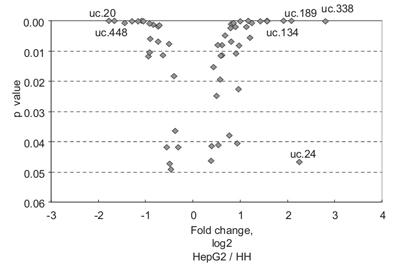
Figure 1. UCR expression is altered in hepatocellular carcinoma. Genome-wide expression profiling in hepatocellular carcinoma and normal liver cells identified 56 UCRS that are inappropriately expressed in malignant hepatocytes. 19 T-UCRs are at least 2-fold up- or downregulated, while 6 (labeled) are more than 3-fold differentially expressed. From Braconi,et al. (2011).
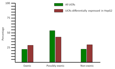
Figure 2. Distribution of aberrantly expressed UCRs in hepatocellular carcinoma. Green bars: All UCRs expressed in the hepatocellular carcinoma cell line HepG2 are distributed according to their subtypes (Exonic, Possibly exonic, Non exonic). Red bars: No dramatic change in the distribution of UCRs according to subtypes is observed for those UCRs that are differentially expressed in HepG2 relative to normal liver. Adapted from Braconi, et al. (2011).
Case study 3: An integrative genomics screen uncovers a correlation of some T-UCRs with clinical prognosis factors in neuroblastoma
Genome-wide expression profiling revealed correlations between specific T-UCR expression levels and important clinicogenetic parameters such as MYCN amplification status in neuroblastoma. Mestdagh, et al. (2010) describe a “signature” of seven T-UCRs that are upregulated in tumors in which MYCN is amplified compared with the less aggressive, MYCN-non-amplified tumors (Figure 3).
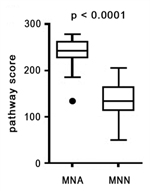
Figure 3. T-UCR expression as a function of MYCN amplification in neuroblastoma. Among 49 patients, the transcriptional levels of seven “signature” T-UCRs are elevated in MYCN-amplified (MNA; left) compared with non-MYCN (MNN) tumors. Ordinate: Pathway score. Adapted from Mestdagh, et al. (2010).
Case Study 4: T-UCR expression as a predictor of survival in neuroblastoma
By comparing 8 short-versus 12 long-term survivors, Scaruffi, et al. (2009) found that the expression levels of 54 T-UCRs are higher in neuroblastoma patients exhibiting long-term (5 years or greater) survival than in short-term survivors, suggesting that T-UCR expression can be used to gauge the prognosis of individuals with this disease. Further, these authors reported that the presence of greater than or fewer than 15 differentially regulated T-UCRs correlated positively with either long- or short-term survival, respectively. While it is not yet clear if the differential expression of any one T-UCR is a causative agent, rather than an effect, of neuroblastoma, this is another example demonstrating that T-UCRs could possibly serve as effective indicators of cancer occurrence, the stage of disease progression, and the chances of a patient’s long-term outlook (Scaruffi, et al., 2009).
Related Services
T-UCR Array Service
LncRNA Array Service
LncRNA qPCR Service
References
Braconi, C., Valeri, N., Kogure, T., Gasparini, P., Huang, N., Nuovo, G.J., Terracciano, L., Croce, C.M., and Patel, T. (2011). Expression and functional role of a transcribed noncoding RNA with an ultraconserved element in hepatocellular carcinoma. Proc Natl Acad Sci U S A 108, 786-791.
Calin, G.A., Liu, C.G., Ferracin, M., Hyslop, T., Spizzo, R., Sevignani, C., Fabbri, M., Cimmino, A., Lee, E.J., Wojcik, S.E., et al. (2007). Ultraconserved regions encoding ncRNAs are altered in human leukemias and carcinomas. Cancer Cell 12, 215-229.
Lujambio, A., Portela, A., Liz, J., Melo, S.A., Rossi, S., Spizzo, R., Croce, C.M., Calin, G.A., and Esteller, M. (2010). CpG island hypermethylation-associated silencing of non-coding RNAs transcribed from ultraconserved regions in human cancer. Oncogene 29, 6390-6401.
Mestdagh, P., Fredlund, E., Pattyn, F., Rihani, A., Van Maerken, T., Vermeulen, J., Kumps, C., Menten, B., De Preter, K., Schramm, A., et al. (2010). An integrative genomics screen uncovers ncRNA T-UCR functions in neuroblastoma tumours. Oncogene 29, 3583-3592.
Scaruffi, P., Stigliani, S., Moretti, S., Coco, S., De Vecchi, C., Valdora, F., Garaventa, A., Bonassi, S., and Tonini, G.P. (2009). Transcribed-Ultra Conserved Region expression is associated with outcome in high-risk neuroblastoma. BMC Cancer 9, 441.