Single-Base RNA Modification Sequencing

Single-Nucleotide RNA m7G Modification Seq(m7G-Quant-seq)

Single-Nucleotide RNA m7G Modification Seq(m7G-Quant-Seq) is an antibody-free, base-resolution method. A targeted chemical conversion distinguishes internal N7-methylguanosine from the 5' cap, detecting and quantifying internal m7G at base resolution.

Arraystar Single-Nucleotide RNA m7G Modification Seq(m7G-Quant-Seq) is an end-to-end sample-to-data service for mRNA, from RNA sample QC and mRNA treatment through library construction, sequencing, and bioinformatics. Sites are annotated to the reference transcriptome with genomic coordinates, host gene, and functional region attribution.

Benefits

In mRNA, internal m7G can influence transcript function, so base-resolution mapping is informative. Targeted conversion separates internal N7-methylguanosine from the cap, detecting and quantifying internal m7G at base resolution.

Single-base resolution: pinpoints m7G sites in mRNA.

Quantitative stoichiometry: conversion-based readouts give accurate modification ratios.

Antibody-free and unbiased: avoids enrichment cross-reactivity and pull-down bias.

Functional annotation: Sites are annotated to the reference transcriptome with genomic coordinates, host gene, and functional region attribution.

Application-ready: supports translation control, RNA stability, coding-region function, and gene-regulation studies across the mRNA transcriptome.

Service NameRNA ClassPrice
Single-Nucleotide RNA m7G Modification Seq(m7G-Quant-Seq)mRNA
Single-Nucleotide RNA m7G Modification Seq(m7G-Quant-Seq)mRNA & lncRNA

Background

In mRNA, the modification shapes transcript stability, translation, and gene regulation, so base-resolution mapping across coding and untranslated regions is what distinguishes it. A targeted chemical conversion distinguishes internal N7-methylguanosine from the 5' cap, recording each internal m7G site as a distinct reverse-transcription signature for base-resolution quantification. This antibody-free readout maps each m7G site at single-base resolution. The same workflow extends to long non-coding RNAs (lncRNAs), enabling researchers to profile this modification on lncRNA transcripts in parallel with mRNA in a single assay.

The method was validated for mRNA, sites are annotated to the reference transcriptome with genomic coordinates, host gene, and functional region attribution. (m7G-quant-seq. ACS Chemical Biology 2022;17(12). PMID: 36398936., DOI: 10.1021/acschembio.2c00792).

Key concept: Single-Nucleotide RNA m7G Modification Seq(m7G-Quant-Seq) is a base-resolution method in which a targeted chemical conversion distinguishes internal n7-methylguanosine from the 5' cap, detecting and quantifying internal m7G at base resolution.
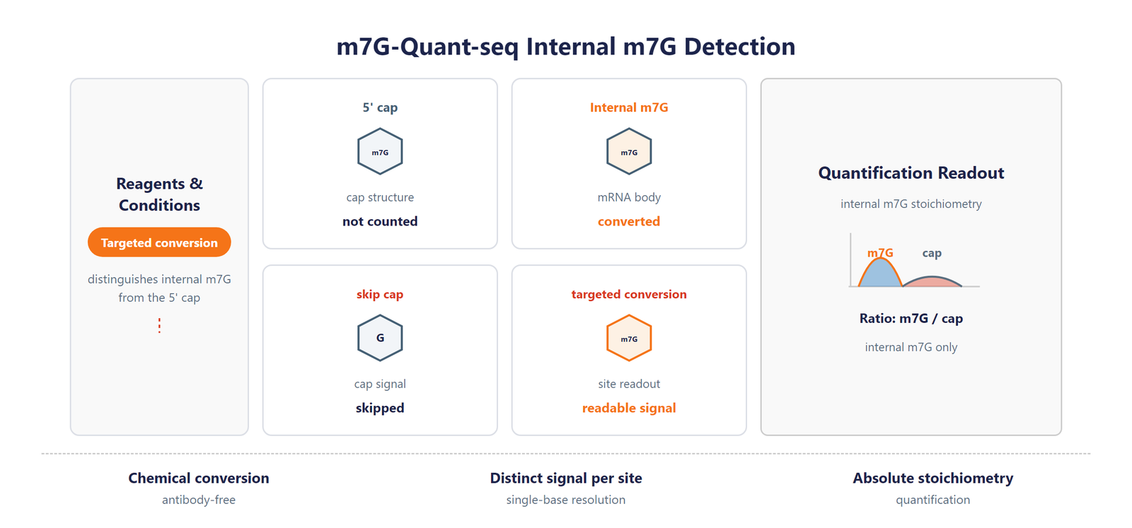
Single-Nucleotide RNA m7G Modification Seq scheme

Figure 1. m7G-Quant-Seq detection scheme. Targeted chemical conversion distinguishes internal N7-methylguanosine from the cap, detecting and quantifying internal m7G at base resolution.

Workflow

Five steps from RNA sample to single-base N7-methylguanosine (m⁷G) data:
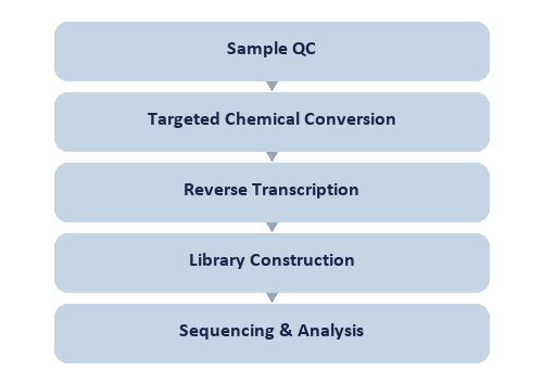
Single-Nucleotide RNA m7G Modification Seq workflow

Figure 2. Single-Nucleotide RNA m7G Modification Seq workflow.

Bioinformatics

The bioinformatics pipeline aligns reads to the reference transcriptome, recognizes m7G-Quant-Seq conversion signatures, and quantifies internal m7G stoichiometry at single-nucleotide resolution, with motif, distribution, differential, enrichment, and genome-browser analyses.

Deliverables

  • Raw m⁷G mRNA sequencing FASTQ data files, formatted for GEO/SRA public database submission.
  • Sample and sequencing QC reports with sorted BAM alignment files and core mapping statistics.
  • High-confidence single-base m⁷G site annotation tables (XLSX) with coordinates, genes, and absolute stoichiometry.
  • Single-base m⁷G transcript feature distribution figures and consensus m⁷G motif logos (PDF/PNG format).
  • Single-base resolution differential m⁷G methylation tables (XLSX) with fold change and significance metrics.
  • Gene Ontology (GO) enrichment analysis reports for m⁷G-modified genes, provided in HTML/PDF/PNG formats.
  • Genome browser-compatible track files (bigWig/bedGraph) and a full structured m⁷G analysis project report.

Research Applications

  • Transcriptome-wide mapping of the modification across protein-coding and non-coding mRNA.
  • Translational control: relates base-resolution modification to mRNA translation and stability.
  • Coding-region function: localizes sites within CDS, UTR, and splice-adjacent regions.
  • Disease-oriented studies: finds differentially modified mRNA sites as biomarkers.

References

[1] m7G-quant-seq: Quantitative Detection of RNA Internal N7-Methylguanosine. ACS Chemical Biology 2022;17(12). DOI: 10.1021/acschembio.2c00792. PMID: 36398936.

Sample Requirements

Storage Guidelines

Cells and Tissues: Preserve in TRIzol or an RNA stabilization solution; snap freeze in liquid nitrogen and store at –80 °C.

RNA: Resuspend in ethanol or RNase-free ultrapure water; store at –80 °C and avoid multiple freeze-thaw cycles.

Shipping Instructions

Place the sample in a 1.5 mL RNase-free microcentrifuge tube.

Seal the tube with parafilm or a cap lock to ensure sample integrity.

Ship the package on dry ice with adequate insulation to maintain the required temperature.

Sample TypeNotes
Whole bloodUse EDTA tubes only, as heparin is not compatible with subsequent analytical procedures.
Cultured cellsSubmission of cell pellets is preferred to ensure high-quality material for processing.
TissueProvide fresh or frozen specimens, ensuring that necrotic material is strictly avoided.
Total RNAMaintain an OD 260/280 ratio ≥ 1.8 and RIN ≥ 7 with no visible degradation. Submit at least 10 µg total RNA.

FAQ

Which mRNA features are annotated?

Sites are annotated to the reference transcriptome with genomic coordinates, host gene, and functional region attribution. This lets you interpret each site in the context of its host gene and mRNA functional region. The method supports both mRNA and tRNA workflows, so you can choose the readout that matches your study.

Can this method quantify the modification at single-base resolution?

Yes. A targeted chemical conversion distinguishes internal N7-methylguanosine from the 5' cap, recording each internal m7G site as a distinct reverse-transcription signature for base-resolution quantification. This provides single-base resolution and absolute stoichiometry for each site in mRNA. The reaction is antibody-free and reproducible, so the calling is specific and the stoichiometry is reliable for downstream functional analyses.

How does this method differ from antibody-based mRNA profiling?

Antibody-based methods rely on enrichment and offer limited resolution with intrinsic motif bias. This method is antibody-free and delivers base-resolution, quantitative calling in mRNA, avoiding cross-reactivity and enrichment bias. This suits projects needing quantitative site-level mapping rather than region-level enrichment, and it pairs well with orthogonal validation.

How does this service support research on m7G modification?

By profiling m7G at base resolution, the service helps researchers explore its roles in RNA function and regulation, providing a valuable evidence base.

Why is high-resolution m7G detection valuable?

Accurate m7G mapping strengthens the evidence behind findings, making it a trusted asset for advancing RNA biology.

Does this service also profile lncRNA?

Yes. We provide two service options: mRNA modification, and mRNA plus long non-coding RNA (lncRNA) modification. Both transcript types can be analyzed together in a single experiment, so there is no need to split them into two projects, and the scope of your study opens up accordingly.

!This assay is provided for research applications only and is not intended for clinical diagnosis, treatment, or personal health evaluation.